Loading...
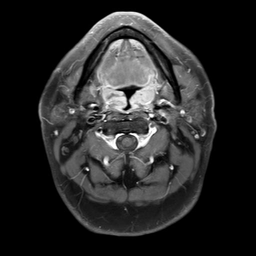
Nasopharyngeal Cancer - Case 005 - 37yo M
58 views6 months agoDiagnosis: Primary Nasopharyngeal Cancer Histologic Type: Non-keratinized squamous cell carcinoma Stage: III (T3N1M0) EBV-DNA Status: (+) This case is from a dataset of patients diagnosed with primary nasopharyngeal carcinoma in The First People’s Hospital of Foshan, China. Three MRI sequences are provided (CE-T1, T1, T2) with corresponding tumor segmentations. By default, the segmentations are disabled. To show them, click on the Setting icon in the bottom right of the DicomTube player and select the "With Segmentations" volume preset. You may also click on the "Tumor Location" display preset to view the tumor with segmentation in all series side by side. License: Creative Commons Attribution 4.0 International (CC BY 4.0) Citation: Li, Y., Chen, Q., & Chen, N. (2024). A dataset of primary nasopharyngeal carcinoma MRI with multi-modalities segmentation [Data set]. Zenodo. https://doi.org/10.5281/zenodo.13131827
NeckMRI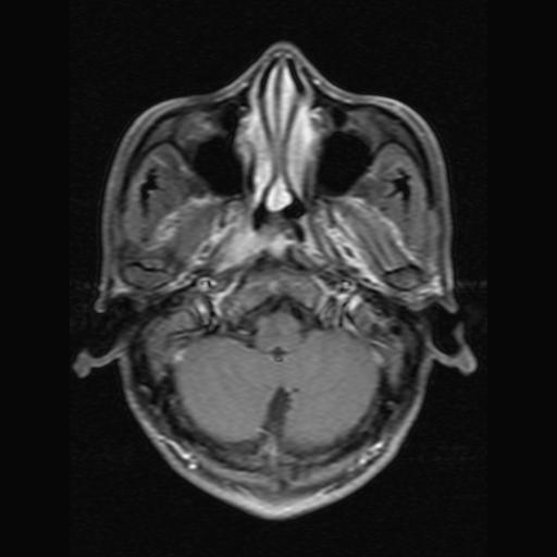
Nasopharyngeal Cancer - Case 004 - 46yo M
72 views6 months agoDiagnosis: Primary Nasopharyngeal Cancer Histologic Type: Non-keratinized squamous cell carcinoma Stage: III (T3N0M0) EBV-DNA Status: (-) This case is from a dataset of patients diagnosed with primary nasopharyngeal carcinoma in The First People’s Hospital of Foshan, China. Three MRI sequences are provided (CE-T1, T1, T2) with corresponding tumor segmentations. By default, the segmentations are disabled. To show them, click on the Setting icon in the bottom right of the DicomTube player and select the "With Segmentations" volume preset. You may also click on the "Tumor Location" display preset to view the tumor with segmentation in all series side by side. License: Creative Commons Attribution 4.0 International (CC BY 4.0) Citation: Li, Y., Chen, Q., & Chen, N. (2024). A dataset of primary nasopharyngeal carcinoma MRI with multi-modalities segmentation [Data set]. Zenodo. https://doi.org/10.5281/zenodo.13131827
NeckMRI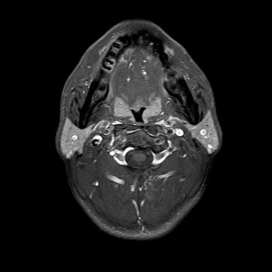
Nasopharyngeal Cancer - Case 003 - 47yo M
79 views6 months agoDiagnosis: Primary Nasopharyngeal Cancer Histologic Type: Non-keratinized squamous cell carcinoma Stage: IVa (T4N1M0) EBV-DNA Status: (-) This case is from a dataset of patients diagnosed with primary nasopharyngeal carcinoma in The First People’s Hospital of Foshan, China. Three MRI sequences are provided (CE-T1, T1, T2) with corresponding tumor segmentations. By default, the segmentations are disabled. To show them, click on the Setting icon in the bottom right of the DicomTube player and select the "With Segmentations" volume preset. You may also click on the "Tumor Location" display preset to view the tumor with segmentation in all series side by side. License: Creative Commons Attribution 4.0 International (CC BY 4.0) Citation: Li, Y., Chen, Q., & Chen, N. (2024). A dataset of primary nasopharyngeal carcinoma MRI with multi-modalities segmentation [Data set]. Zenodo. https://doi.org/10.5281/zenodo.13131827
NeckMRI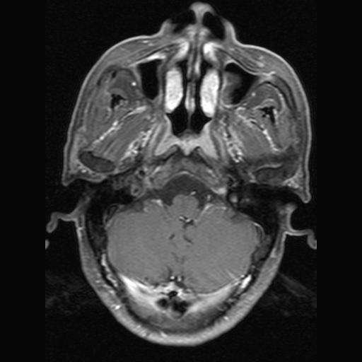
Nasopharyngeal Cancer - Case 002 - 60yo M
72 views6 months agoDiagnosis: Primary Nasopharyngeal Cancer Histologic Type: Non-keratinized squamous cell carcinoma Stage: IVa (T2N3M0) EBV-DNA Status: (-) This case is from a dataset of patients diagnosed with primary nasopharyngeal carcinoma in The First People’s Hospital of Foshan, China. Three MRI sequences are provided (CE-T1, T1, T2) with corresponding tumor segmentations. By default, the segmentations are disabled. To show them, click on the Setting icon in the bottom right of the DicomTube player and select the "With Segmentations" volume preset. You may also click on the "Tumor Location" display preset to view the tumor with segmentation in all series side by side. License: Creative Commons Attribution 4.0 International (CC BY 4.0) Citation: Li, Y., Chen, Q., & Chen, N. (2024). A dataset of primary nasopharyngeal carcinoma MRI with multi-modalities segmentation [Data set]. Zenodo. https://doi.org/10.5281/zenodo.13131827
NeckMRI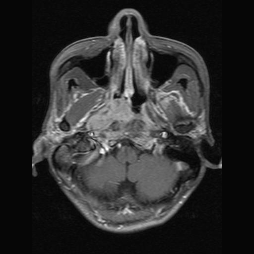
Nasopharyngeal Cancer - Case 001 - 55yo M
90 views6 months agoDiagnosis: Primary Nasopharyngeal Cancer Histologic Type: Non-keratinized squamous cell carcinoma Stage: III (T3N2M0) EBV-DNA Status: (+) This case is from a dataset of patients diagnosed with primary nasopharyngeal carcinoma in The First People’s Hospital of Foshan, China. Three MRI sequences are provided (CE-T1, T1, T2) with corresponding tumor segmentations. By default, the segmentations are disabled. To show them, click on the Setting icon in the bottom right of the DicomTube player and select the "With Segmentations" volume preset. You may also click on the "Tumor Location" display preset to view the tumor with segmentation in all series side by side. License: Creative Commons Attribution 4.0 International (CC BY 4.0) Citation: Li, Y., Chen, Q., & Chen, N. (2024). A dataset of primary nasopharyngeal carcinoma MRI with multi-modalities segmentation [Data set]. Zenodo. https://doi.org/10.5281/zenodo.13131827
NeckMRI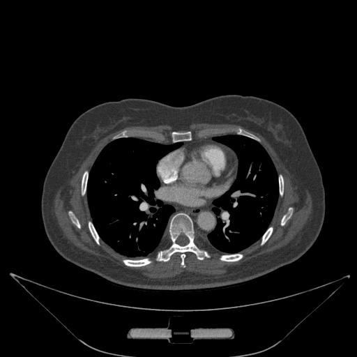
Tetralogy Of Fallot - ImageCHD - Case 1178
22 views6 months agoDiagnosis: tetralogy of Fallot This case is from ImageCHD, a 3D Computed Tomography (CT) image dataset for classification of Congenital Heart Disease (CHD). The dataset includes 110 CT images with cardiac segmentations covering most types of CHD. By default, the cardiac segmentations are disabled. To show them, click on the Setting icon in the bottom right of the DicomTube player and select the "With Segmentations" volume preset. Cardiac Segmentation Labels: Left Ventricle (red) Right Ventricle (green) Left Atrium (blue) Right Atrium (yellow) Myocardium (magenta) Aorta (cyan) Pulmonary Artery (orange) License: Apache 2.0 Citation: Xiaowei Xu, Tianchen Wang, Haiyun Yuan, Qianjun Jia, Jianzheng Ceng, Yuhao Dong, Meiping Huang, Jian Zhuang, Yiyu Shi, "ImageCHD: A 3D Computed Tomography Image Dataset for Classification of Congenital Heart Disease," in Proc. of Medical Image Computing and Computer Assisted Interventions (MICCAI), Online, 2020.
ChestCT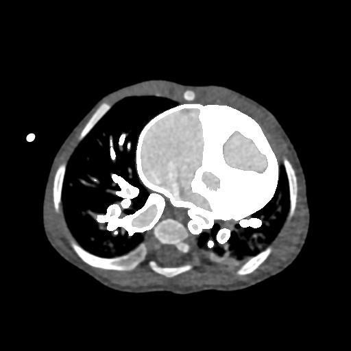
Atrial Septal Defect, Anomalous Pulmonary Venous Drainage - ImageCHD - Case 1170
8 views6 months agoDiagnosis: atrial septal defect, anomalous pulmonary venous drainage This case is from ImageCHD, a 3D Computed Tomography (CT) image dataset for classification of Congenital Heart Disease (CHD). The dataset includes 110 CT images with cardiac segmentations covering most types of CHD. By default, the cardiac segmentations are disabled. To show them, click on the Setting icon in the bottom right of the DicomTube player and select the "With Segmentations" volume preset. Cardiac Segmentation Labels: Left Ventricle (red) Right Ventricle (green) Left Atrium (blue) Right Atrium (yellow) Myocardium (magenta) Aorta (cyan) Pulmonary Artery (orange) License: Apache 2.0 Citation: Xiaowei Xu, Tianchen Wang, Haiyun Yuan, Qianjun Jia, Jianzheng Ceng, Yuhao Dong, Meiping Huang, Jian Zhuang, Yiyu Shi, "ImageCHD: A 3D Computed Tomography Image Dataset for Classification of Congenital Heart Disease," in Proc. of Medical Image Computing and Computer Assisted Interventions (MICCAI), Online, 2020.
ChestCT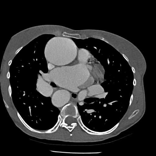
Atrial Septal Defect, Ventricular Septal Defect, Pulmonary Atresia, Double Superior Vena Cava - ImageCHD - Case 1161
10 views6 months agoDiagnosis: atrial septal defect, ventricular septal defect, pulmonary atresia, double superior vena cava This case is from ImageCHD, a 3D Computed Tomography (CT) image dataset for classification of Congenital Heart Disease (CHD). The dataset includes 110 CT images with cardiac segmentations covering most types of CHD. By default, the cardiac segmentations are disabled. To show them, click on the Setting icon in the bottom right of the DicomTube player and select the "With Segmentations" volume preset. Cardiac Segmentation Labels: Left Ventricle (red) Right Ventricle (green) Left Atrium (blue) Right Atrium (yellow) Myocardium (magenta) Aorta (cyan) Pulmonary Artery (orange) License: Apache 2.0 Citation: Xiaowei Xu, Tianchen Wang, Haiyun Yuan, Qianjun Jia, Jianzheng Ceng, Yuhao Dong, Meiping Huang, Jian Zhuang, Yiyu Shi, "ImageCHD: A 3D Computed Tomography Image Dataset for Classification of Congenital Heart Disease," in Proc. of Medical Image Computing and Computer Assisted Interventions (MICCAI), Online, 2020.
ChestCT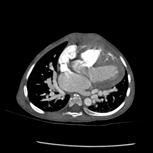
Ventricular Septal Defect, Pulmonary Atresia - ImageCHD - Case 1158
9 views6 months agoDiagnosis: ventricular septal defect, pulmonary atresia This case is from ImageCHD, a 3D Computed Tomography (CT) image dataset for classification of Congenital Heart Disease (CHD). The dataset includes 110 CT images with cardiac segmentations covering most types of CHD. By default, the cardiac segmentations are disabled. To show them, click on the Setting icon in the bottom right of the DicomTube player and select the "With Segmentations" volume preset. Cardiac Segmentation Labels: Left Ventricle (red) Right Ventricle (green) Left Atrium (blue) Right Atrium (yellow) Myocardium (magenta) Aorta (cyan) Pulmonary Artery (orange) License: Apache 2.0 Citation: Xiaowei Xu, Tianchen Wang, Haiyun Yuan, Qianjun Jia, Jianzheng Ceng, Yuhao Dong, Meiping Huang, Jian Zhuang, Yiyu Shi, "ImageCHD: A 3D Computed Tomography Image Dataset for Classification of Congenital Heart Disease," in Proc. of Medical Image Computing and Computer Assisted Interventions (MICCAI), Online, 2020.
ChestCT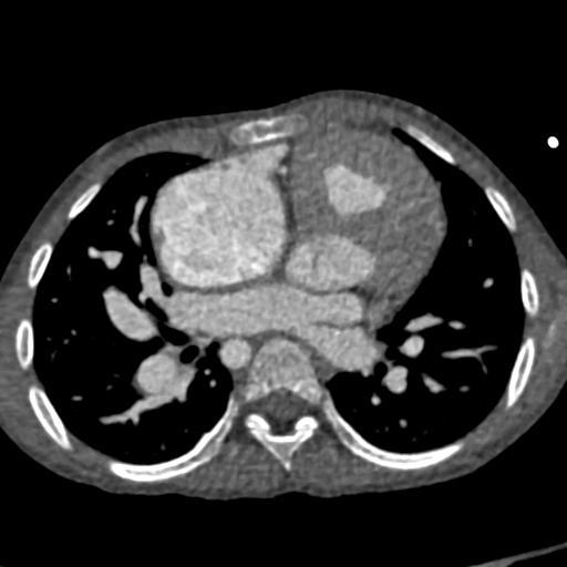
Pulmonary Atresia - ImageCHD - Case 1150
6 views6 months agoDiagnosis: pulmonary atresia This case is from ImageCHD, a 3D Computed Tomography (CT) image dataset for classification of Congenital Heart Disease (CHD). The dataset includes 110 CT images with cardiac segmentations covering most types of CHD. By default, the cardiac segmentations are disabled. To show them, click on the Setting icon in the bottom right of the DicomTube player and select the "With Segmentations" volume preset. Cardiac Segmentation Labels: Left Ventricle (red) Right Ventricle (green) Left Atrium (blue) Right Atrium (yellow) Myocardium (magenta) Aorta (cyan) Pulmonary Artery (orange) License: Apache 2.0 Citation: Xiaowei Xu, Tianchen Wang, Haiyun Yuan, Qianjun Jia, Jianzheng Ceng, Yuhao Dong, Meiping Huang, Jian Zhuang, Yiyu Shi, "ImageCHD: A 3D Computed Tomography Image Dataset for Classification of Congenital Heart Disease," in Proc. of Medical Image Computing and Computer Assisted Interventions (MICCAI), Online, 2020.
ChestCT