Loading...
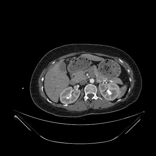
Type-A Aortic Dissection from Image-TAAD (8)
69 views6 months agoDiagnosis: Type A Aortic Dissection This case is from Image-TAAD, a dataset providing comprehensive segmentation of Type-A aortic dissection (TAAD). The segmentations include true lumen (TL), false lumen (FL), intimal tear, aortic branches, and surrounding organs. By default, the aortic dissection segmentations are disabled. To show them, click on the Setting icon in the bottom right of the DicomTube player and select the "With Segmentations" volume preset. License: Apache 2.0 Citation: Shanshan Song, Hailong Qiu, Meiping Huang, Jian Zhuang, Qing Lu, Yiyu Shi, Xiaomeng Li, Wen Xie, Guang Tong, Xiaowei Xu, "Domain knowledge based comprehensive segmentation of Type-A aortic dissection with clinically-oriented evaluation," Medical Image Analysis, Volume 102, 2025, 103512. https://doi.org/10.1016/j.media.2025.103512
CAPCT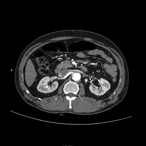
Type-A Aortic Dissection from Image-TAAD (7)
67 views6 months agoDiagnosis: Type A Aortic Dissection This case is from Image-TAAD, a dataset providing comprehensive segmentation of Type-A aortic dissection (TAAD). The segmentations include true lumen (TL), false lumen (FL), intimal tear, aortic branches, and surrounding organs. By default, the aortic dissection segmentations are disabled. To show them, click on the Setting icon in the bottom right of the DicomTube player and select the "With Segmentations" volume preset. License: Apache 2.0 Citation: Shanshan Song, Hailong Qiu, Meiping Huang, Jian Zhuang, Qing Lu, Yiyu Shi, Xiaomeng Li, Wen Xie, Guang Tong, Xiaowei Xu, "Domain knowledge based comprehensive segmentation of Type-A aortic dissection with clinically-oriented evaluation," Medical Image Analysis, Volume 102, 2025, 103512. https://doi.org/10.1016/j.media.2025.103512
CAPCT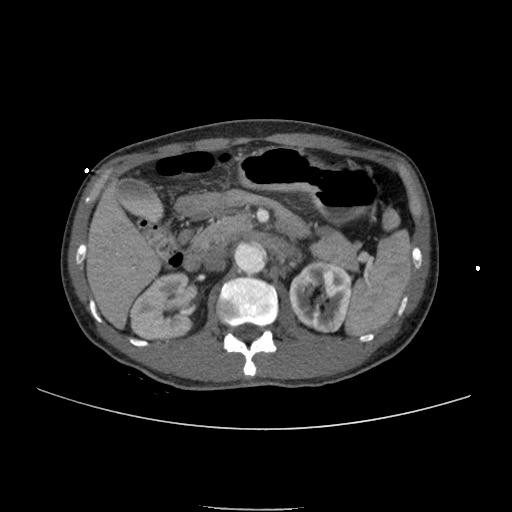
Type-A Aortic Dissection from Image-TAAD (6)
72 views6 months agoDiagnosis: Type A Aortic Dissection This case is from Image-TAAD, a dataset providing comprehensive segmentation of Type-A aortic dissection (TAAD). The segmentations include true lumen (TL), false lumen (FL), intimal tear, aortic branches, and surrounding organs. By default, the aortic dissection segmentations are disabled. To show them, click on the Setting icon in the bottom right of the DicomTube player and select the "With Segmentations" volume preset. License: Apache 2.0 Citation: Shanshan Song, Hailong Qiu, Meiping Huang, Jian Zhuang, Qing Lu, Yiyu Shi, Xiaomeng Li, Wen Xie, Guang Tong, Xiaowei Xu, "Domain knowledge based comprehensive segmentation of Type-A aortic dissection with clinically-oriented evaluation," Medical Image Analysis, Volume 102, 2025, 103512. https://doi.org/10.1016/j.media.2025.103512
CAPCT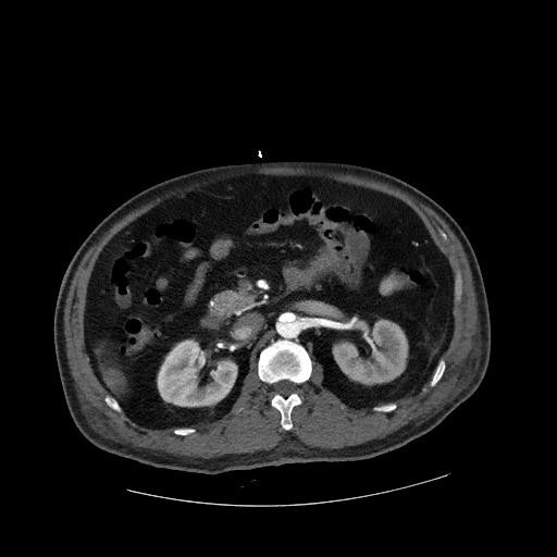
Type-A Aortic Dissection from Image-TAAD (5)
71 views6 months agoDiagnosis: Type A Aortic Dissection This case is from Image-TAAD, a dataset providing comprehensive segmentation of Type-A aortic dissection (TAAD). The segmentations include true lumen (TL), false lumen (FL), intimal tear, aortic branches, and surrounding organs. By default, the aortic dissection segmentations are disabled. To show them, click on the Setting icon in the bottom right of the DicomTube player and select the "With Segmentations" volume preset. License: Apache 2.0 Citation: Shanshan Song, Hailong Qiu, Meiping Huang, Jian Zhuang, Qing Lu, Yiyu Shi, Xiaomeng Li, Wen Xie, Guang Tong, Xiaowei Xu, "Domain knowledge based comprehensive segmentation of Type-A aortic dissection with clinically-oriented evaluation," Medical Image Analysis, Volume 102, 2025, 103512. https://doi.org/10.1016/j.media.2025.103512
CAPCT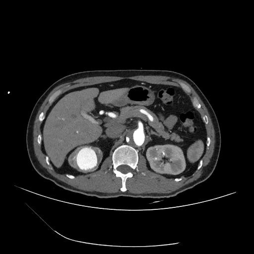
Type-A Aortic Dissection from Image-TAAD (4)
89 views6 months agoDiagnosis: Type A Aortic Dissection This case is from Image-TAAD, a dataset providing comprehensive segmentation of Type-A aortic dissection (TAAD). The segmentations include true lumen (TL), false lumen (FL), intimal tear, aortic branches, and surrounding organs. By default, the aortic dissection segmentations are disabled. To show them, click on the Setting icon in the bottom right of the DicomTube player and select the "With Segmentations" volume preset. License: Apache 2.0 Citation: Shanshan Song, Hailong Qiu, Meiping Huang, Jian Zhuang, Qing Lu, Yiyu Shi, Xiaomeng Li, Wen Xie, Guang Tong, Xiaowei Xu, "Domain knowledge based comprehensive segmentation of Type-A aortic dissection with clinically-oriented evaluation," Medical Image Analysis, Volume 102, 2025, 103512. https://doi.org/10.1016/j.media.2025.103512
CAPCT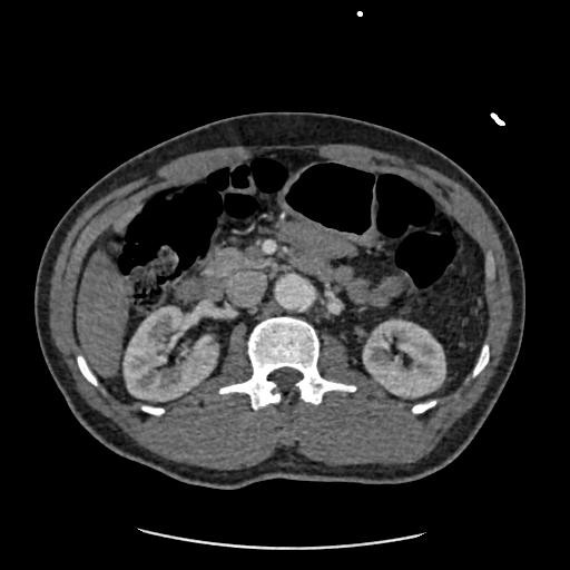
Type-A Aortic Dissection from Image-TAAD (2)
115 views6 months agoDiagnosis: Type A Aortic Dissection This case is from Image-TAAD, a dataset providing comprehensive segmentation of Type-A aortic dissection (TAAD). The segmentations include true lumen (TL), false lumen (FL), intimal tear, aortic branches, and surrounding organs. By default, the aortic dissection segmentations are disabled. To show them, click on the Setting icon in the bottom right of the DicomTube player and select the "With Segmentations" volume preset. License: Apache 2.0 Citation: Shanshan Song, Hailong Qiu, Meiping Huang, Jian Zhuang, Qing Lu, Yiyu Shi, Xiaomeng Li, Wen Xie, Guang Tong, Xiaowei Xu, "Domain knowledge based comprehensive segmentation of Type-A aortic dissection with clinically-oriented evaluation," Medical Image Analysis, Volume 102, 2025, 103512. https://doi.org/10.1016/j.media.2025.103512
CAPCT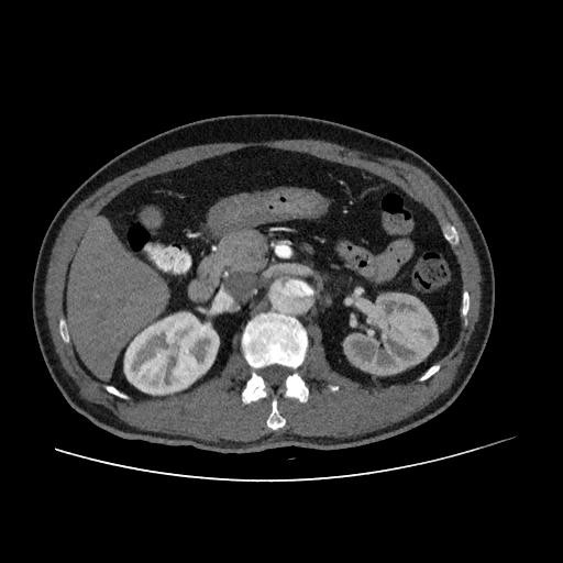
Type-A Aortic Dissection from Image-TAAD (1)
121 views6 months agoDiagnosis: Type A Aortic Dissection This case is from Image-TAAD, a dataset providing comprehensive segmentation of Type-A aortic dissection (TAAD). The segmentations include true lumen (TL), false lumen (FL), intimal tear, aortic branches, and surrounding organs. By default, the aortic dissection segmentations are disabled. To show them, click on the Setting icon in the bottom right of the DicomTube player and select the "With Segmentations" volume preset. License: Apache 2.0 Citation: Shanshan Song, Hailong Qiu, Meiping Huang, Jian Zhuang, Qing Lu, Yiyu Shi, Xiaomeng Li, Wen Xie, Guang Tong, Xiaowei Xu, "Domain knowledge based comprehensive segmentation of Type-A aortic dissection with clinically-oriented evaluation," Medical Image Analysis, Volume 102, 2025, 103512. https://doi.org/10.1016/j.media.2025.103512
CAPCT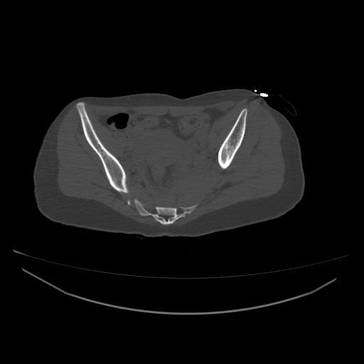
Pelvic Fracture - PENGWIN 100
68 views6 months agoThis case is from the PENGWIN Task 1 dataset, which contains 150 CT scans of patients scheduled for pelvic reduction surgery due to pelvic fractures. The dataset includes segmentations for sacrum and hipbone fragments, which have been semi-automatically annotated and validated by medical experts. Each bone anatomy (sacrum, left hipbone, right hipbone) has up to 10 fragments labeled. Bone without fracture is represented as a single fragment. IMPORTANT NOTE: Based on my review of a handfull of the segmentations, not all fractures are accurately labelled, especially minimally displaced fractures. Use segmentations with caution. In the future, we may manually annotate these images to better delineate all fractures and provide standardized characterization of the fracture patterns. By default, the fracture segmentation is disabled. To show it, click on the Settings icon in the bottom right of the DicomTube player and select the "With Segmentation" volume preset. License: Creative Commons Attribution 4.0 International (CC BY 4.0) Citation: Sang, Y., Liu, Y., Yibulayimu, S., Zhu, G., Wang, Y., Killeen, B., Liu, M., Ku, P.-C., Armand, M., Unberath, M., Wu, X., & Zhao, C. (2024). PENGWIN Task 1: Pelvic Fracture Segmentation on CT. Zenodo. https://doi.org/10.5281/zenodo.10927452
PelvisCT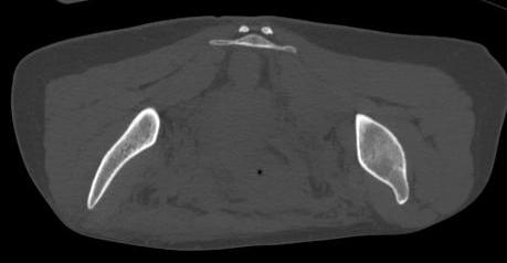
Pelvic Fracture - PENGWIN 099
18 views6 months agoThis case is from the PENGWIN Task 1 dataset, which contains 150 CT scans of patients scheduled for pelvic reduction surgery due to pelvic fractures. The dataset includes segmentations for sacrum and hipbone fragments, which have been semi-automatically annotated and validated by medical experts. Each bone anatomy (sacrum, left hipbone, right hipbone) has up to 10 fragments labeled. Bone without fracture is represented as a single fragment. IMPORTANT NOTE: Based on my review of a handfull of the segmentations, not all fractures are accurately labelled, especially minimally displaced fractures. Use segmentations with caution. In the future, we may manually annotate these images to better delineate all fractures and provide standardized characterization of the fracture patterns. By default, the fracture segmentation is disabled. To show it, click on the Settings icon in the bottom right of the DicomTube player and select the "With Segmentation" volume preset. License: Creative Commons Attribution 4.0 International (CC BY 4.0) Citation: Sang, Y., Liu, Y., Yibulayimu, S., Zhu, G., Wang, Y., Killeen, B., Liu, M., Ku, P.-C., Armand, M., Unberath, M., Wu, X., & Zhao, C. (2024). PENGWIN Task 1: Pelvic Fracture Segmentation on CT. Zenodo. https://doi.org/10.5281/zenodo.10927452
PelvisCT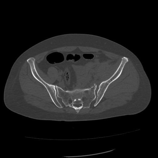
Pelvic Fracture - PENGWIN 098
15 views6 months agoThis case is from the PENGWIN Task 1 dataset, which contains 150 CT scans of patients scheduled for pelvic reduction surgery due to pelvic fractures. The dataset includes segmentations for sacrum and hipbone fragments, which have been semi-automatically annotated and validated by medical experts. Each bone anatomy (sacrum, left hipbone, right hipbone) has up to 10 fragments labeled. Bone without fracture is represented as a single fragment. IMPORTANT NOTE: Based on my review of a handfull of the segmentations, not all fractures are accurately labelled, especially minimally displaced fractures. Use segmentations with caution. In the future, we may manually annotate these images to better delineate all fractures and provide standardized characterization of the fracture patterns. By default, the fracture segmentation is disabled. To show it, click on the Settings icon in the bottom right of the DicomTube player and select the "With Segmentation" volume preset. License: Creative Commons Attribution 4.0 International (CC BY 4.0) Citation: Sang, Y., Liu, Y., Yibulayimu, S., Zhu, G., Wang, Y., Killeen, B., Liu, M., Ku, P.-C., Armand, M., Unberath, M., Wu, X., & Zhao, C. (2024). PENGWIN Task 1: Pelvic Fracture Segmentation on CT. Zenodo. https://doi.org/10.5281/zenodo.10927452
PelvisCT