Loading...
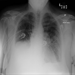
Atelectasis (from the NIH Chest X-Ray dataset)
2 views3 months ago@Key findings demonstrate Atelectasis (red bounding box). This case is taken from the NIH Chest X-Ray dataset provided by the NIH Clinical Center. The findings above are taken directly from the metadata in the dataset and are not independently verified. License: No restrictions on use as long as you provide the link to the original download site, acknowledge the NIH Clinical Center, and provide a citation to the CVPR 2017 paper below. Original download link: https://nihcc.app.box.com/v/ChestXray-NIHCC Citation: X. Wang, Y. Peng, L. Lu, Z. Lu, M. Bagheri and R. M. Summers, "ChestX-Ray8: Hospital-Scale Chest X-Ray Database and Benchmarks on Weakly-Supervised Classification and Localization of Common Thorax Diseases," 2017 IEEE Conference on Computer Vision and Pattern Recognition (CVPR), Honolulu, HI, USA, 2017, pp. 3462-3471, doi: 10.1109/CVPR.2017.369.
ChestXR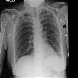
Pneumonia (from the NIH Chest X-Ray dataset)
27 views3 months ago@Key findings demonstrate Pneumonia (rose bounding box). This case is taken from the NIH Chest X-Ray dataset provided by the NIH Clinical Center. The findings above are taken directly from the metadata in the dataset and are not independently verified. License: No restrictions on use as long as you provide the link to the original download site, acknowledge the NIH Clinical Center, and provide a citation to the CVPR 2017 paper below. Original download link: https://nihcc.app.box.com/v/ChestXray-NIHCC Citation: X. Wang, Y. Peng, L. Lu, Z. Lu, M. Bagheri and R. M. Summers, "ChestX-Ray8: Hospital-Scale Chest X-Ray Database and Benchmarks on Weakly-Supervised Classification and Localization of Common Thorax Diseases," 2017 IEEE Conference on Computer Vision and Pattern Recognition (CVPR), Honolulu, HI, USA, 2017, pp. 3462-3471, doi: 10.1109/CVPR.2017.369.
ChestXR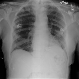
Atelectasis (from the NIH Chest X-Ray dataset)
6 views3 months ago@Key findings demonstrate Atelectasis (red bounding box). This case is taken from the NIH Chest X-Ray dataset provided by the NIH Clinical Center. The findings above are taken directly from the metadata in the dataset and are not independently verified. License: No restrictions on use as long as you provide the link to the original download site, acknowledge the NIH Clinical Center, and provide a citation to the CVPR 2017 paper below. Original download link: https://nihcc.app.box.com/v/ChestXray-NIHCC Citation: X. Wang, Y. Peng, L. Lu, Z. Lu, M. Bagheri and R. M. Summers, "ChestX-Ray8: Hospital-Scale Chest X-Ray Database and Benchmarks on Weakly-Supervised Classification and Localization of Common Thorax Diseases," 2017 IEEE Conference on Computer Vision and Pattern Recognition (CVPR), Honolulu, HI, USA, 2017, pp. 3462-3471, doi: 10.1109/CVPR.2017.369.
ChestXR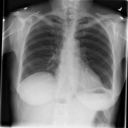
Atelectasis (from the NIH Chest X-Ray dataset)
3 views3 months ago@Key findings demonstrate Atelectasis (red bounding box). This case is taken from the NIH Chest X-Ray dataset provided by the NIH Clinical Center. The findings above are taken directly from the metadata in the dataset and are not independently verified. License: No restrictions on use as long as you provide the link to the original download site, acknowledge the NIH Clinical Center, and provide a citation to the CVPR 2017 paper below. Original download link: https://nihcc.app.box.com/v/ChestXray-NIHCC Citation: X. Wang, Y. Peng, L. Lu, Z. Lu, M. Bagheri and R. M. Summers, "ChestX-Ray8: Hospital-Scale Chest X-Ray Database and Benchmarks on Weakly-Supervised Classification and Localization of Common Thorax Diseases," 2017 IEEE Conference on Computer Vision and Pattern Recognition (CVPR), Honolulu, HI, USA, 2017, pp. 3462-3471, doi: 10.1109/CVPR.2017.369.
ChestXR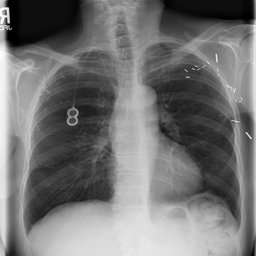
Atelectasis (from the NIH Chest X-Ray dataset)
6 views3 months ago@Key findings demonstrate Atelectasis (red bounding box). This case is taken from the NIH Chest X-Ray dataset provided by the NIH Clinical Center. The findings above are taken directly from the metadata in the dataset and are not independently verified. License: No restrictions on use as long as you provide the link to the original download site, acknowledge the NIH Clinical Center, and provide a citation to the CVPR 2017 paper below. Original download link: https://nihcc.app.box.com/v/ChestXray-NIHCC Citation: X. Wang, Y. Peng, L. Lu, Z. Lu, M. Bagheri and R. M. Summers, "ChestX-Ray8: Hospital-Scale Chest X-Ray Database and Benchmarks on Weakly-Supervised Classification and Localization of Common Thorax Diseases," 2017 IEEE Conference on Computer Vision and Pattern Recognition (CVPR), Honolulu, HI, USA, 2017, pp. 3462-3471, doi: 10.1109/CVPR.2017.369.
ChestXR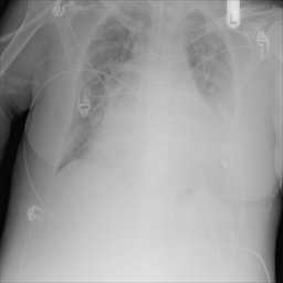
Infiltrate (from the NIH Chest X-Ray dataset)
4 views3 months ago@Key findings demonstrate Infiltrate (orange bounding box). This case is taken from the NIH Chest X-Ray dataset provided by the NIH Clinical Center. The findings above are taken directly from the metadata in the dataset and are not independently verified. License: No restrictions on use as long as you provide the link to the original download site, acknowledge the NIH Clinical Center, and provide a citation to the CVPR 2017 paper below. Original download link: https://nihcc.app.box.com/v/ChestXray-NIHCC Citation: X. Wang, Y. Peng, L. Lu, Z. Lu, M. Bagheri and R. M. Summers, "ChestX-Ray8: Hospital-Scale Chest X-Ray Database and Benchmarks on Weakly-Supervised Classification and Localization of Common Thorax Diseases," 2017 IEEE Conference on Computer Vision and Pattern Recognition (CVPR), Honolulu, HI, USA, 2017, pp. 3462-3471, doi: 10.1109/CVPR.2017.369.
ChestXR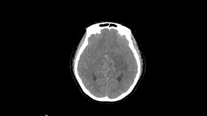
Germinoma - Case 067
105 views4 months agoDiagnosis: Germinoma This case includes CT imaging with corresponding tumor segmentations. By default, the segmentations are disabled. To show them, click on the Setting icon in the bottom right of the DicomTube player and select the "With Segmentations" volume preset. You may also click on the Tumor Location display preset to view the tumor with segmentation. License: Creative Commons Attribution 4.0 International (CC BY 4.0) Citation: Huang, Lixuan; Jiangnian, Gong; Liang, Lun (2025). A comprehensive dataset of germinoma on MRI/CT with clinical and radiomic data. figshare. Dataset. https://doi.org/10.6084/m9.figshare.28045148.v1
BrainCT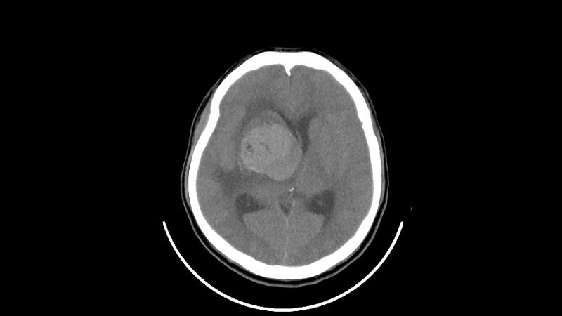
Germinoma - Case 066
86 views4 months agoDiagnosis: Germinoma This case includes CT imaging with corresponding tumor segmentations. By default, the segmentations are disabled. To show them, click on the Setting icon in the bottom right of the DicomTube player and select the "With Segmentations" volume preset. You may also click on the Tumor Location display preset to view the tumor with segmentation. License: Creative Commons Attribution 4.0 International (CC BY 4.0) Citation: Huang, Lixuan; Jiangnian, Gong; Liang, Lun (2025). A comprehensive dataset of germinoma on MRI/CT with clinical and radiomic data. figshare. Dataset. https://doi.org/10.6084/m9.figshare.28045148.v1
BrainCT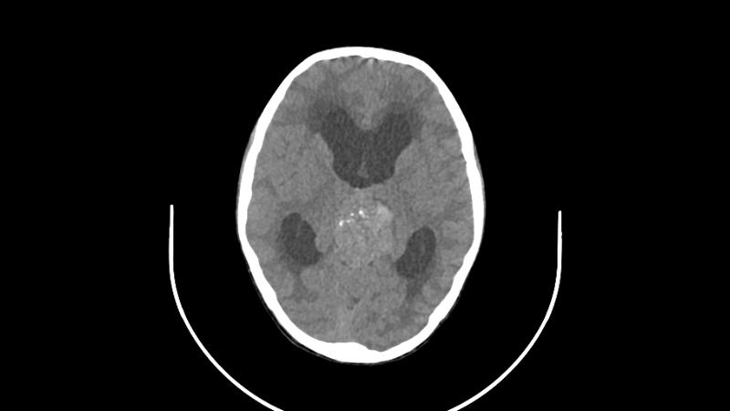
Germinoma - Case 065
66 views4 months agoDiagnosis: Germinoma This case includes CT imaging with corresponding tumor segmentations. By default, the segmentations are disabled. To show them, click on the Setting icon in the bottom right of the DicomTube player and select the "With Segmentations" volume preset. You may also click on the Tumor Location display preset to view the tumor with segmentation. License: Creative Commons Attribution 4.0 International (CC BY 4.0) Citation: Huang, Lixuan; Jiangnian, Gong; Liang, Lun (2025). A comprehensive dataset of germinoma on MRI/CT with clinical and radiomic data. figshare. Dataset. https://doi.org/10.6084/m9.figshare.28045148.v1
BrainCT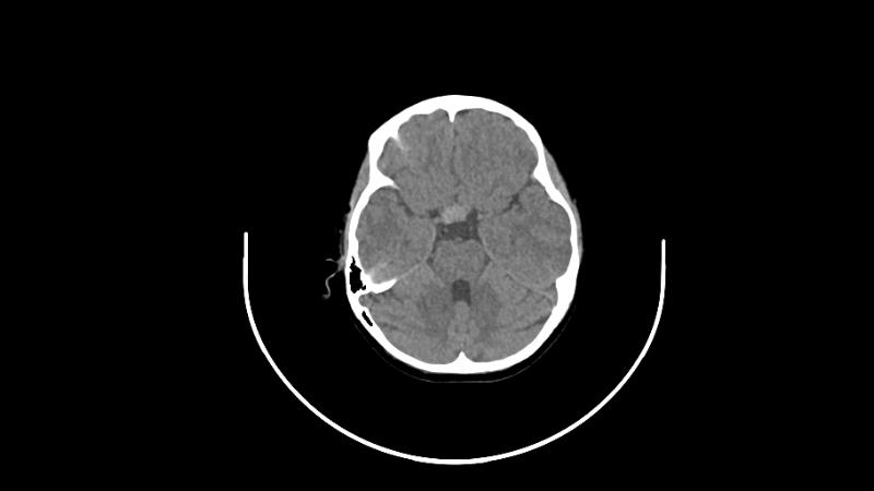
Germinoma - Case 064
59 views4 months agoDiagnosis: Germinoma This case includes CT imaging with corresponding tumor segmentations. By default, the segmentations are disabled. To show them, click on the Setting icon in the bottom right of the DicomTube player and select the "With Segmentations" volume preset. You may also click on the Tumor Location display preset to view the tumor with segmentation. License: Creative Commons Attribution 4.0 International (CC BY 4.0) Citation: Huang, Lixuan; Jiangnian, Gong; Liang, Lun (2025). A comprehensive dataset of germinoma on MRI/CT with clinical and radiomic data. figshare. Dataset. https://doi.org/10.6084/m9.figshare.28045148.v1
BrainCT