Loading...
Anatomy Modules
13 cases
Updated 6 months ago
EEDNeuroRad
A collection of interactive 3D anatomy modules utilizing the DicomTube platform.
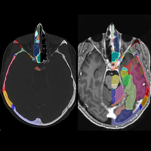
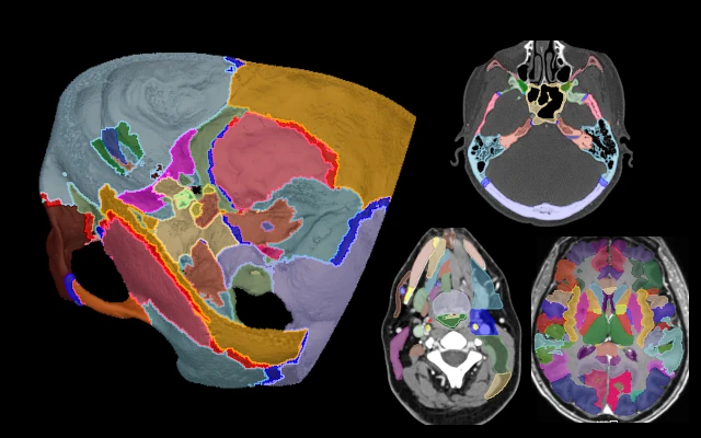
Comprehensive Head CT + MRI Anatomy: Brain, Skullbase and Sinonasal Cavity
3,453 views
6 months ago
Highly detailed annotated anatomy of the skull, skullbase, brain, and sinonasal cavity on registered CT and MRI images from the same subject. There are multiple display presets available which you can access from the settings button in the bottom right of the DicomTube player.The brain cortical segmentation is predominantly auto generated using Freesurfer software and the Desikan-Killiany Atlas. For further details visit the Freesurfer website or associated publication. There are, however, a number of manual edits and custom post processing also performed on the cortical segmentations to make sure they respect the anatomy better.
Head
MRI, CT
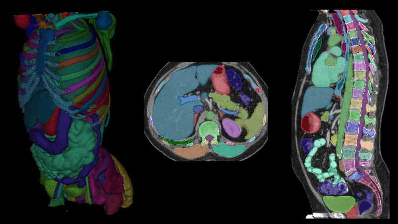
Body CT Anatomy Module from Total Segmentator Dataset (s0011)
2,059 views
6 months ago
Case and segmentations are taken directly from the Total Segmentator dataset, which is distributed under the CC Attribution 4.0 International license. If interested, you may display a default 3D view of the segmentations via the settings button in the bottom right corner of the DicomTube player.The following additional metadata are provided for this particular case in the dataset:Pathology: tumor Pathology Location: thorax,abdomen,pelvisCitation:Jakob Wasserthal. (2023). Dataset with segmentations of 117 important anatomical structures in 1228 CT images (2.0.1) [Data set]. Zenodo. https://doi.org/10.5281/zenodo.10047292
Whole Body
CT

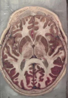
Head Anatomy Module from the Visible Human Project (Half Resolution)
372 views
about 1 month ago
Additional head images from the Visible Human Project, courtesy of the U.S. National Library of Medicine. For these images, the donor was preserved in formalin and the blood vessels were filled with araldite-F. After freezing the specimen was cryo sectioned at 0.147mm intervals and digital photographs were taken with a resolution of 1056 x 1528 pixels. This case has been downsampled by a factor of 2 from the originals for decreased file sizes and to have the ability for 3D processing. Automatic brain cortical and subcortical segmentations are performed using the Freesurfer software, in particular the SynthSeg program (for details see SynthSeg: Segmentation of brain MRI scans of any contrast and resolution without retraining. B Billot, DN Greve, O Puonti, A Thielscher, K Van Leemput, B Fischl, AV Dalca, JE Iglesias. Medical Image Analysis, 83, 102789 (2023)). The cortical segmentations were subsequently expanded for improved visibility. You can use the keyboard shortcut "a" to toggle the visibility of the segmentations.
Head
Cryo
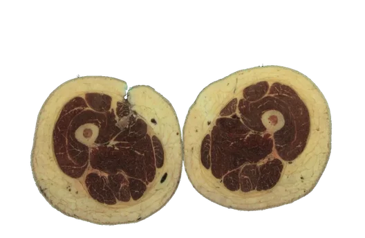
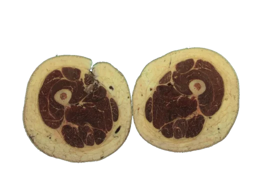
Thigh Anatomy Module from the Visible Human Project (Half Resolution)
117 views
26 days ago
Anatomy module made from the thigh portion of the female images from the Visible Human Project, courtesy of the U.S. National Library of Medicine. The anatomic segmentations were performed by researchers at the University of Denver and the Center for Orthopaedic Biomechanics with citation below. You may press the keyboard shortcut 'a' to toggle visibility of the color segmentations. The original cryo cross-section images were at 0.33 mm intervals and with each pixel 0.33 mm in size. This case has been downsampled in the z-axis by a factor of 6 and in the x and y-axis by a factor 2 to allow 3D processing. License: Creative Commons Attribution 4.0 International License (CC BY 4.0) for the segmentations Citations: T. E. Andreassen, D. R. Hume, L. D. Hamilton, K. E. Walker, S. E. Higinbotham, and K. B. Shelburne, “Three Dimensional Lower Extremity Musculoskeletal Geometry of the Visible Human Female and Male,” Sci. Data, vol. 10, no. 1, p. 34, Jan. 2023, doi: 10.1038/s41597-022-01905-2.
Thigh
Cryo

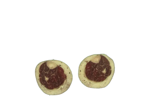
Leg Anatomy Module from the Visible Human Project (Half Resolution)
40 views
26 days ago
Anatomy module made from the leg portion of the female images from the Visible Human Project, courtesy of the U.S. National Library of Medicine. The anatomic segmentations were performed by researchers at the University of Denver and the Center for Orthopaedic Biomechanics with citation below. You may press the keyboard shortcut 'a' to toggle visibility of the color segmentations. The original cryo cross-section images were at 0.33 mm intervals and with each pixel 0.33 mm in size. This case has been downsampled in the z-axis by a factor of 6 and in the x and y-axis by a factor 2 to allow 3D processing. License: Creative Commons Attribution 4.0 International License (CC BY 4.0) for the segmentations Citations: T. E. Andreassen, D. R. Hume, L. D. Hamilton, K. E. Walker, S. E. Higinbotham, and K. B. Shelburne, “Three Dimensional Lower Extremity Musculoskeletal Geometry of the Visible Human Female and Male,” Sci. Data, vol. 10, no. 1, p. 34, Jan. 2023, doi: 10.1038/s41597-022-01905-2.
Leg
Cryo
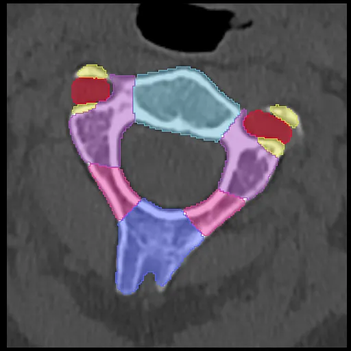
Cervical Spine Anatomy
548 views
6 months ago
Interactive 3D anatomy module of the cervical spine with labeled structures.
Cervical Spine
CT
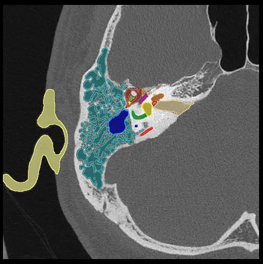
Temporal Bone Anatomy
781 views
6 months ago
Interactive 3D anatomy module of the temporal bone with labeled structures.
Temporal Bone
CT
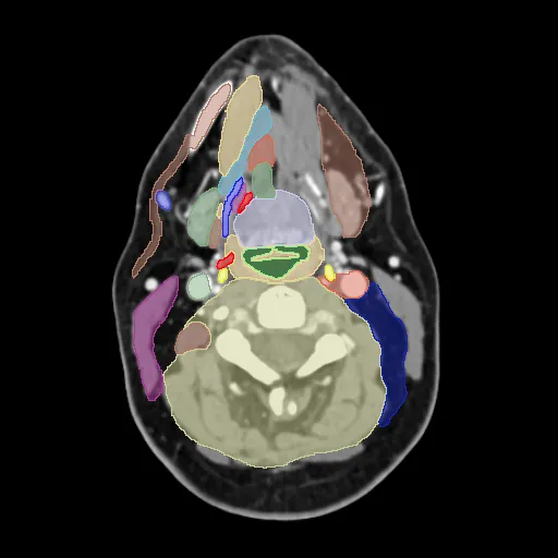
Neck CT Anatomy
1,193 views
6 months ago
Interactive 3D anatomy module of the neck with labeled structures and deep spaces.
Neck
CT

Intracranial Vessel Anatomy (CTA) from TOP-BRAIN Dataset (004)
325 views
6 months ago
Case and segmentations are taken directly from the TOP-BRAIN dataset (TopBrain Segmentation Challenge for Whole Brain Vessel Anatomy), which is distributed under a license requiring source attribution. This dataset contains voxel annotations for over 40 landmark brain vessel anatomies covering both arteries and veins.If interested, you may display a default 3D view of the segmentations via the settings button in the bottom right corner of the DicomTube player.License: opendata.swiss Open use. Must provide the source. Use for commercial purposes requires permission of the data owner.Citation:TopBrain Challenge: TopBrain Challenge Organizers. (2025). TopBrain Challenge Data Release [Data set]. Zenodo. https://doi.org/10.5281/zenodo.16878417TopCoW Challenge pre-print: Benchmarking the CoW with the TopCoW Challenge: Topology-Aware Anatomical Segmentation of the Circle of Willis for CTA and MRA. Kaiyuan Yang et. al. 2025. arXiv 2312.17670
Head
CT
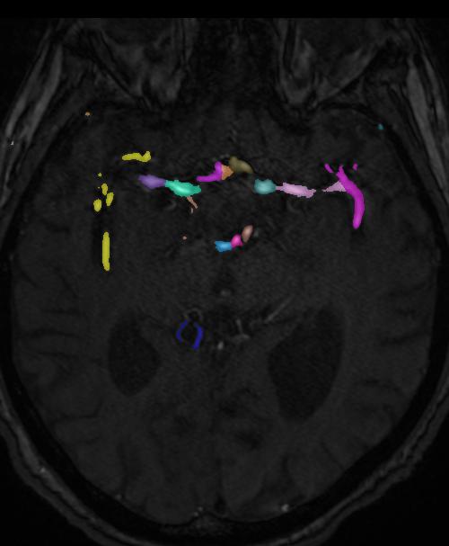
Intracranial Vessel Anatomy (MRA) from TOP-BRAIN Dataset (004)
213 views
6 months ago
Case and segmentations are taken directly from the TOP-BRAIN dataset (TopBrain Segmentation Challenge for Whole Brain Vessel Anatomy), which is distributed under a license requiring source attribution. This dataset contains voxel annotations for over 40 landmark brain vessel anatomies covering both arteries and veins.If interested, you may display a default 3D view of the segmentations via the settings button in the bottom right corner of the DicomTube player.License: opendata.swiss Open use. Must provide the source. Use for commercial purposes requires permission of the data owner.Citation:TopBrain Challenge: TopBrain Challenge Organizers. (2025). TopBrain Challenge Data Release [Data set]. Zenodo. https://doi.org/10.5281/zenodo.16878417TopCoW Challenge pre-print: Benchmarking the CoW with the TopCoW Challenge: Topology-Aware Anatomical Segmentation of the Circle of Willis for CTA and MRA. Kaiyuan Yang et. al. 2025. arXiv 2312.17670
Head
MR
Head Anatomy Module from the Visible Human Project
122 views
about 1 month ago
Additional head images from the Visible Human Project, courtesy of the U.S. National Library of Medicine. For these images, the donor was preserved in formalin and the blood vessels were filled with araldite-F. After freezing the specimen was cryo sectioned at 0.147mm intervals and digital photographs were taken with a resolution of 1056 x 1528 pixels. Automatic brain cortical and subcortical segmentations are performed using the Freesurfer software, in particular the SynthSeg program (for details see SynthSeg: Segmentation of brain MRI scans of any contrast and resolution without retraining. B Billot, DN Greve, O Puonti, A Thielscher, K Van Leemput, B Fischl, AV Dalca, JE Iglesias. Medical Image Analysis, 83, 102789 (2023)). The cortical segmentations were subsequently expanded for improved visibility. You can use the keyboard shortcut "a" to toggle the visibility of the segmentations. Note: Due to the large file size, attempting to perform either 3D MPR or 3D VRT on this dataset will crash your current tab's WebGL session. This would then require a full page refresh to be functional again. If you need to do 3D processing, use the "half resoltion" variant instead.
Head
Cryo
Thigh Anatomy Module from the Visible Human Project
34 views
26 days ago
Anatomy module made from the thigh portion of the female images from the Visible Human Project, courtesy of the U.S. National Library of Medicine. The anatomic segmentations were performed by researchers at the University of Denver and the Center for Orthopaedic Biomechanics with citation below. You may press the keyboard shortcut 'a' to toggle visibility of the color segmentations. The original cryo cross-section images were at 0.33 mm intervals and with each pixel 0.33 mm in size. In order to be able to fit the dataset in memory, this case has been downsampled in the z-axis by a factor of 3. The cryo sections are therefore at 1 mm intervals, similar to the male dataset. Note: Due to the large file size, attempting to perform either 3D MPR or 3D VRT on this dataset will crash your current tab's WebGL session. This would then require a full page refresh to be functional again. If you need to do 3D processing, use the "half resoltion" variant instead. License: Creative Commons Attribution 4.0 International License (CC BY 4.0) for the segmentations Citations: T. E. Andreassen, D. R. Hume, L. D. Hamilton, K. E. Walker, S. E. Higinbotham, and K. B. Shelburne, “Three Dimensional Lower Extremity Musculoskeletal Geometry of the Visible Human Female and Male,” Sci. Data, vol. 10, no. 1, p. 34, Jan. 2023, doi: 10.1038/s41597-022-01905-2.
Thigh
Cryo
Leg Anatomy Module from the Visible Human Project
39 views
26 days ago
Anatomy module made from the leg portion of the female images from the Visible Human Project, courtesy of the U.S. National Library of Medicine. The anatomic segmentations were performed by researchers at the University of Denver and the Center for Orthopaedic Biomechanics with citation below. You may press the keyboard shortcut 'a' to toggle visibility of the color segmentations. The original cryo cross-section images were at 0.33 mm intervals and with each pixel 0.33 mm in size. In order to be able to fit the dataset in memory, this case has been downsampled in the z-axis by a factor of 3. The cryo sections are therefore at 1 mm intervals, similar to the male dataset. Note: Due to the large file size, attempting to perform either 3D MPR or 3D VRT on this dataset will crash your current tab's WebGL session. This would then require a full page refresh to be functional again. If you need to do 3D processing, use the "half resoltion" variant instead. License: Creative Commons Attribution 4.0 International License (CC BY 4.0) for the segmentations Citations: T. E. Andreassen, D. R. Hume, L. D. Hamilton, K. E. Walker, S. E. Higinbotham, and K. B. Shelburne, “Three Dimensional Lower Extremity Musculoskeletal Geometry of the Visible Human Female and Male,” Sci. Data, vol. 10, no. 1, p. 34, Jan. 2023, doi: 10.1038/s41597-022-01905-2.
Leg
Cryo