Loading...
Latest Cases
Discover the latest published medical cases from our community
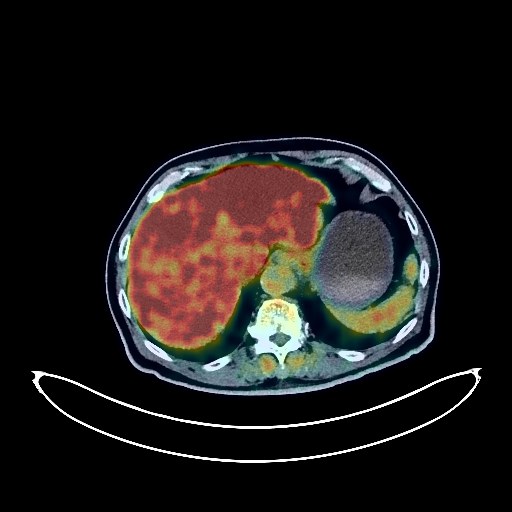
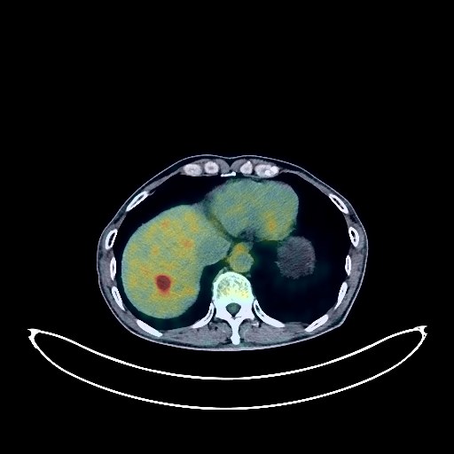
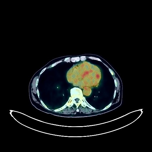
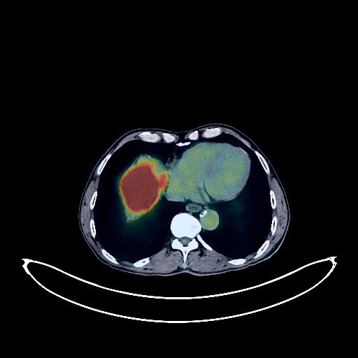
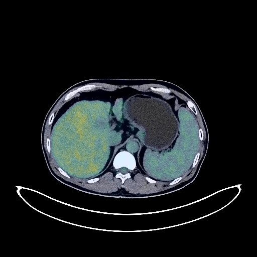
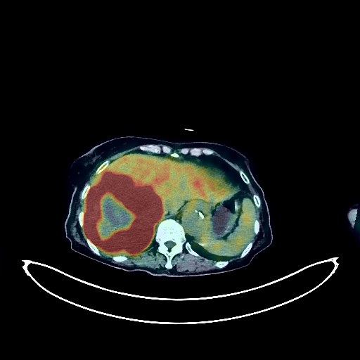
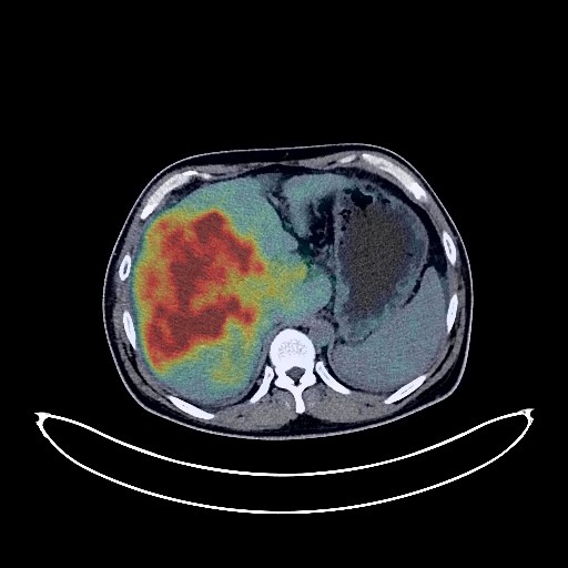
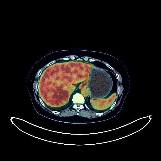
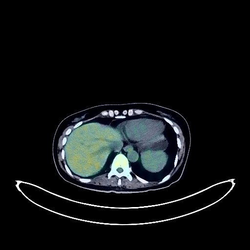
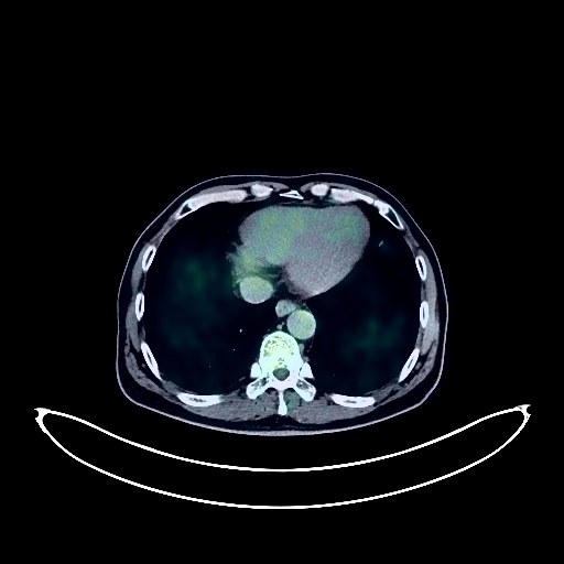
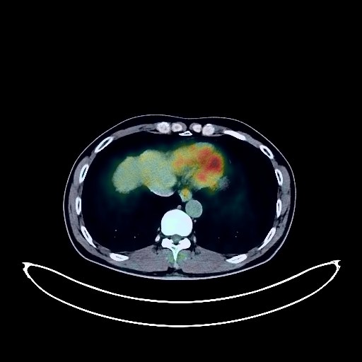
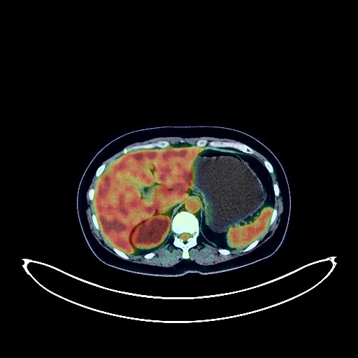
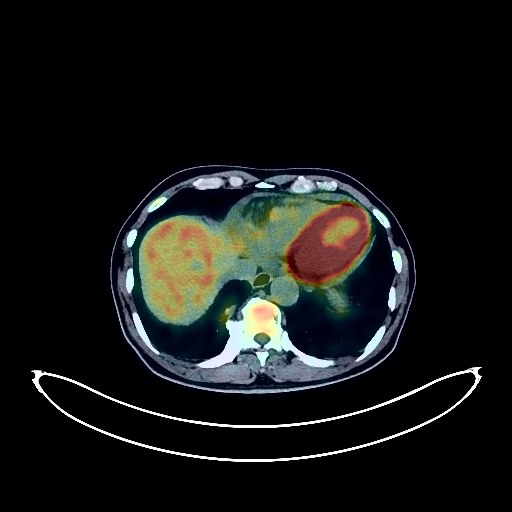
Liver Cancer PET/CT (case 984005-000027 from PETWB-REP)
17 views
9 days ago
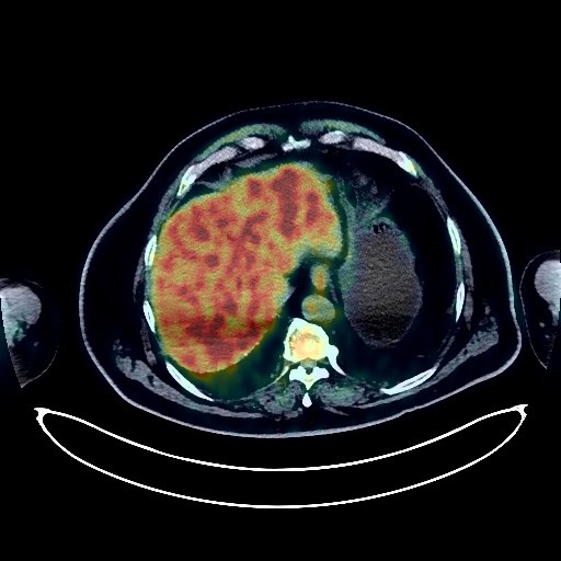
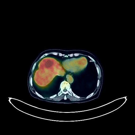
Whole-body 18F-FDG PET/CT scan in a patient with Liver Cancer taken from the PETWB-REP dataset. The following English report (translated from original Chinese) is taken verbatim from the public dataset and has not been modified or otherwise checked for accuracy (see the end for citation). Impression a. A slightly low-density mass in liver S6 with unevenly increased FDG metabolism. Based on the MRI report from another hospital, malignancy is suspected, with liver cancer being the primary consideration. Please confirm with pathology. b. Several lymph nodes in the hepatic hilum, hilar space, and retroperitoneum are shown, with normal FDG metabolism. Reactive lymph node hyperplasia is suspected. Please follow up. a. Pure ground-glass nodules in both lungs with normal FDG metabolism are suspected, possibly due to chronic inflammation or atypical adenomatous hyperplasia. Please have an annual CT scan. b. Small (solid) chronic inflammatory nodules in both lungs. Please follow up with CT scan. A few post-inflammatory lesions in both lungs. Mild thickening of the pleura on both sides. Calcification of some arterial walls. Calcification of the prostate. Dilation of both inguinal canals. Increased FDG metabolism in parts of the stomach wall and intestines, possibly due to physiological metabolism or chronic inflammation. Please have an endoscopy. Osteoporosis. Spinal degeneration, bilateral pars interarticularis fracture of L5 vertebral body with grade I anterior spondylolisthesis. Bilateral deep lacunar infarcts, age-related brain changes. Minor inflammation of bilateral maxillary sinuses. This case is from PETWB-REP, a curated dataset of whole-body 18F-FDG PET/CT scans and corresponding radiology reports from 490 patients with a broad spectrum of malignancies. The data were retrospectively collected from patients who underwent clinically indicated whole-body 18F-FDG PET/CT scans at the Shanghai Universal Medical Imaging Diagnostic Center between 2021 and 2024. License: Creative Commons Attribution 4.0 International (CC BY 4.0) Citation: Xue, L., Feng, G., Wenbo, Z., Zhang, Y., Li, L., Wang, S., Peng, L., Peng, S., & Gao, X. (2026). PETWB-REP: A Multi-Cancer Whole-Body FDG PET/CT Dataset with Corresponding Radiology Reports [Data set]. Zenodo. https://doi.org/10.5281/zenodo.18670487
Whole Body
PET/CT
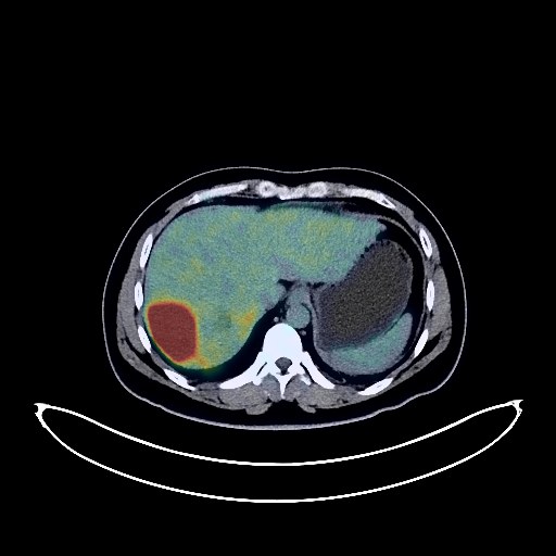
Liver Cancer PET/CT (case 984005-000026 from PETWB-REP)
6 views
9 days ago
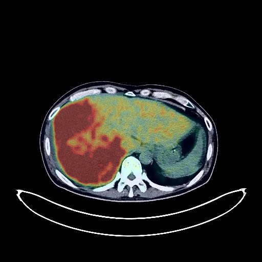
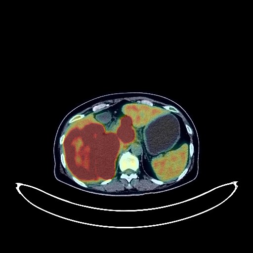
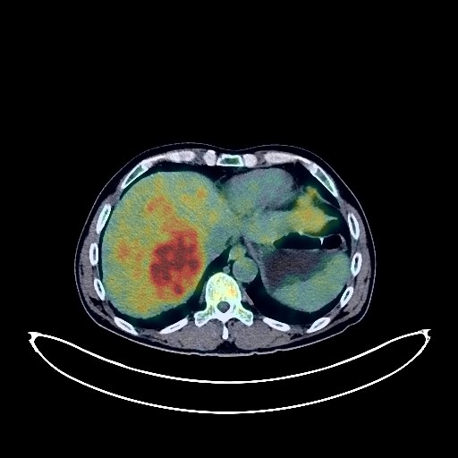
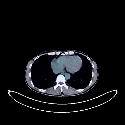
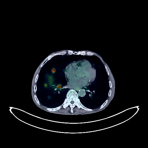
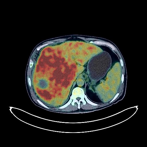
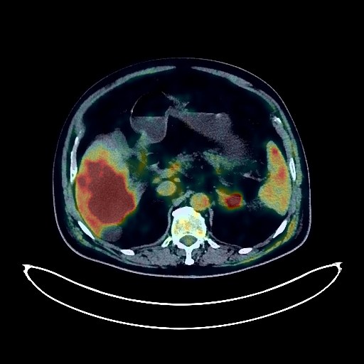
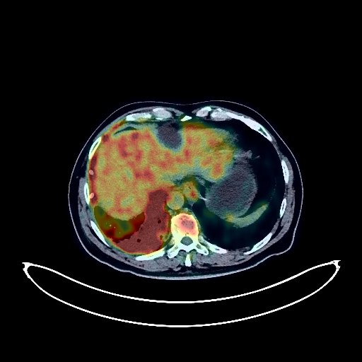
Whole-body 18F-FDG PET/CT scan in a patient with Liver Cancer taken from the PETWB-REP dataset. The following English report (translated from original Chinese) is taken verbatim from the public dataset and has not been modified or otherwise checked for accuracy (see the end for citation). Impression a. Large, slightly low-density masses in the right lobe and left inner lobe of the liver (partially nodular fusion-like changes) with increased FDG metabolism, suggestive of hepatocellular carcinoma with intrahepatic metastasis. b. Slight thickening of the right branch and main trunk of the portal vein with increased FDG metabolism, suggestive of tumor thrombus formation. c. Liver cirrhosis. Cholestasis of the gallbladder. Abdominal and pelvic effusions. a. Mixed ground-glass opacities in the anterior segment of the right upper lobe of the lung, with mildly increased FDG metabolism, suggestive of inflammatory lesions, atypical lung cancer to be ruled out, CT scan recommended in 1-3 months. b. Scattered chronic inflammatory nodules in both lungs are the primary consideration, scattered inflammation in both lungs (partially chronic inflammation and sequelae), CT scan recommended after anti-inflammatory treatment. c. Mild thickening of the pleura on both sides. Tracheal diverticulum. Calcification of the wall of part of the left branch of the coronary artery. The right adrenal gland is poorly visualized, and the left adrenal gland shows mild hyperplasia. Degenerative changes in the spine. L4/5 and L5/S1 disc herniation, the latter with partial calcification and pneumoconiosis. Cranial scintigraphy showed no obvious abnormalities. Minor inflammation of the left maxillary sinus. This case is from PETWB-REP, a curated dataset of whole-body 18F-FDG PET/CT scans and corresponding radiology reports from 490 patients with a broad spectrum of malignancies. The data were retrospectively collected from patients who underwent clinically indicated whole-body 18F-FDG PET/CT scans at the Shanghai Universal Medical Imaging Diagnostic Center between 2021 and 2024. License: Creative Commons Attribution 4.0 International (CC BY 4.0) Citation: Xue, L., Feng, G., Wenbo, Z., Zhang, Y., Li, L., Wang, S., Peng, L., Peng, S., & Gao, X. (2026). PETWB-REP: A Multi-Cancer Whole-Body FDG PET/CT Dataset with Corresponding Radiology Reports [Data set]. Zenodo. https://doi.org/10.5281/zenodo.18670487
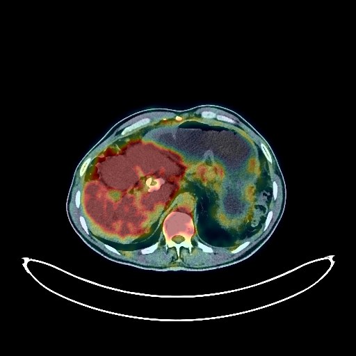
Liver Cancer PET/CT (case 984005-000025 from PETWB-REP)
7 views
9 days ago
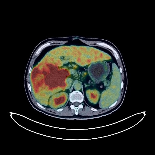
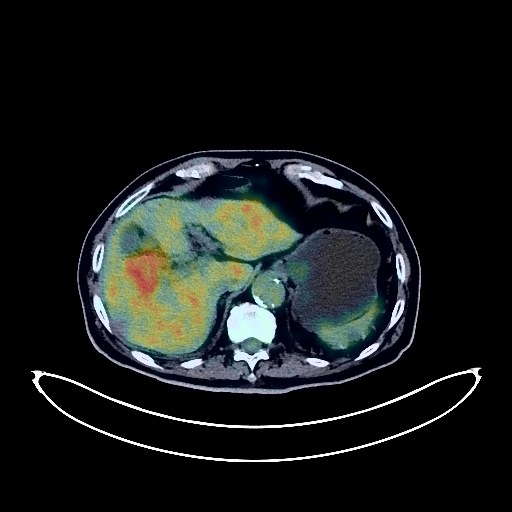
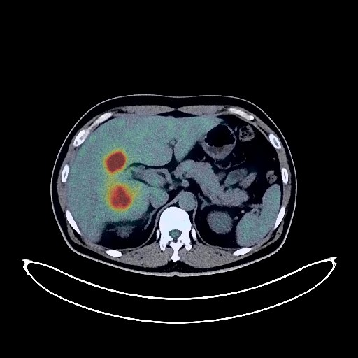
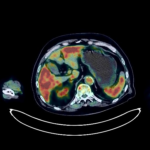
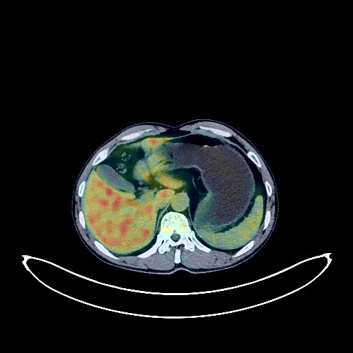
Whole-body 18F-FDG PET/CT scan in a patient with Liver Cancer taken from the PETWB-REP dataset. The following English report (translated from original Chinese) is taken verbatim from the public dataset and has not been modified or otherwise checked for accuracy (see the end for citation). Impression a. Irregular, slightly low-density nodules and masses in the right lobe of the liver, with increased FDG metabolism, suggestive of hepatocellular carcinoma (highly likely) with intrahepatic lesions or metastatic tumor formation. b. Tumor thrombus formation in the right branch of the portal vein and the inferior vena cava. Partial lymph node metastasis in the hepatogastric space, hepatic hilum, and hilar space needs to be ruled out; close observation is recommended. c. Liver cirrhosis. Slightly enlarged spleen. Multiple chronic inflammatory micronodules and calcifications in both lungs; please follow up with CT to rule out mixed metastatic lesions. A few post-inflammatory lesions in both lungs. Partial arteriosclerosis. Slightly increased FDG metabolism in the gastric wall, suggestive of physiological metabolism or chronic inflammatory changes; please follow up with endoscopy. Degenerative changes in the spine. L4/5 and L5/S1 intervertebral disc bulges. No obvious abnormalities were found on cranial scintigraphy. This case is from PETWB-REP, a curated dataset of whole-body 18F-FDG PET/CT scans and corresponding radiology reports from 490 patients with a broad spectrum of malignancies. The data were retrospectively collected from patients who underwent clinically indicated whole-body 18F-FDG PET/CT scans at the Shanghai Universal Medical Imaging Diagnostic Center between 2021 and 2024. License: Creative Commons Attribution 4.0 International (CC BY 4.0) Citation: Xue, L., Feng, G., Wenbo, Z., Zhang, Y., Li, L., Wang, S., Peng, L., Peng, S., & Gao, X. (2026). PETWB-REP: A Multi-Cancer Whole-Body FDG PET/CT Dataset with Corresponding Radiology Reports [Data set]. Zenodo. https://doi.org/10.5281/zenodo.18670487
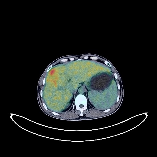
Liver Cancer PET/CT (case 984005-000024 from PETWB-REP)
5 views
9 days ago
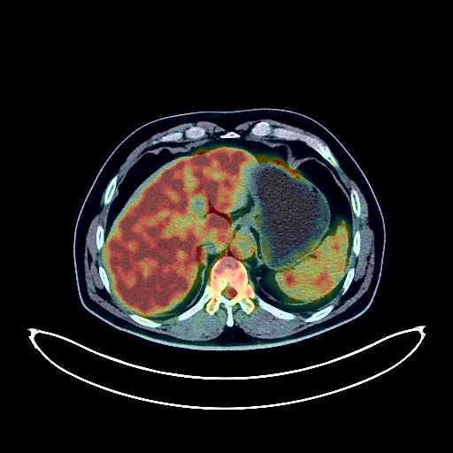
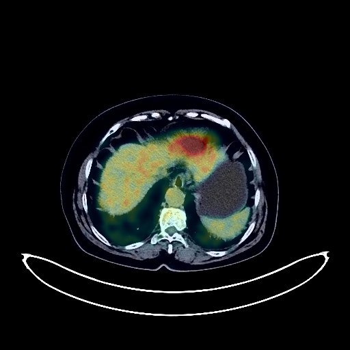
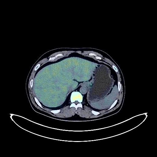
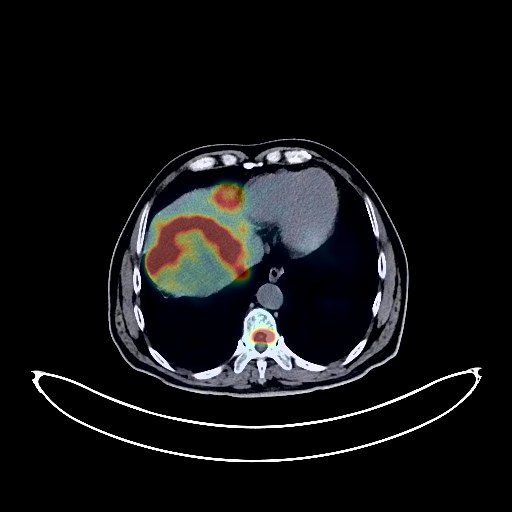
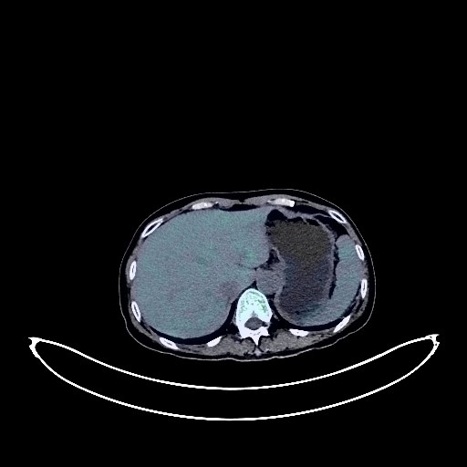
Whole-body 18F-FDG PET/CT scan in a patient with Liver Cancer taken from the PETWB-REP dataset. The following English report (translated from original Chinese) is taken verbatim from the public dataset and has not been modified or otherwise checked for accuracy (see the end for citation). Impression a. Slightly low-density nodules with increased FDG metabolism in the lower segment of the right lobe of the liver, suggestive of malignancy, such as cholangiocarcinoma or poorly differentiated hepatocellular carcinoma. Please confirm with pathology. b. Several small cysts in the liver. Chronic inflammatory micronodules in both lungs. Please confirm with CT follow-up. A few post-inflammatory lesions in both lungs. Absent after cholecystectomy. Accessory spleen. Mild hyperplasia of the left adrenal gland. Small cysts in both kidneys. Calcifications in the prostate. Increased FDG metabolism in the stomach wall, colon, and part of the rectum, suggestive of physiological metabolism or chronic inflammatory changes. Please confirm with endoscopy follow-up. Degenerative changes in the spine. Schmorl's nodes at the lower margins of the T7 and L4 vertebral bodies. L4/5 and L5/S1 intervertebral disc bulges. No obvious abnormalities were found on cranial scintigraphy. A few inflammations in the right sphenoid sinus and bilateral ethmoid sinuses. This case is from PETWB-REP, a curated dataset of whole-body 18F-FDG PET/CT scans and corresponding radiology reports from 490 patients with a broad spectrum of malignancies. The data were retrospectively collected from patients who underwent clinically indicated whole-body 18F-FDG PET/CT scans at the Shanghai Universal Medical Imaging Diagnostic Center between 2021 and 2024. License: Creative Commons Attribution 4.0 International (CC BY 4.0) Citation: Xue, L., Feng, G., Wenbo, Z., Zhang, Y., Li, L., Wang, S., Peng, L., Peng, S., & Gao, X. (2026). PETWB-REP: A Multi-Cancer Whole-Body FDG PET/CT Dataset with Corresponding Radiology Reports [Data set]. Zenodo. https://doi.org/10.5281/zenodo.18670487
Liver Cancer PET/CT (case 984005-000023 from PETWB-REP)
6 views
9 days ago
Whole-body 18F-FDG PET/CT scan in a patient with Liver Cancer taken from the PETWB-REP dataset. The following English report (translated from original Chinese) is taken verbatim from the public dataset and has not been modified or otherwise checked for accuracy (see the end for citation). Impression a. A mass in the left lobe of the liver with increased FDG metabolism, suggestive of hepatocellular carcinoma; several slightly low-density lesions in the remaining liver, with FDG showing background metabolism, metastatic or daughter lesions are the primary consideration, please combine with enhanced MRI for comprehensive analysis. Reactive hyperplasia of the hilar and retroperitoneal lymph nodes. b. Multiple metastatic tumors in both lungs. Chronic inflammation and sequelae in both lungs. Bilateral pleural thickening. Postoperative changes after cardiac surgery, calcification of some arterial walls (including coronary arteries). Chronic cholecystitis, gallstones. Left adrenal hyperplasia. Left renal cyst. Prostatic hyperplasia with calcification. Chronic inflammatory changes or physiological metabolism in some intestinal segments, please combine with endoscopic follow-up. Osteoporosis, degenerative changes in the spine. Multiple lumbar disc herniations with pneumothorax and degeneration. Age-related brain changes, deep lacunar infarcts. This case is from PETWB-REP, a curated dataset of whole-body 18F-FDG PET/CT scans and corresponding radiology reports from 490 patients with a broad spectrum of malignancies. The data were retrospectively collected from patients who underwent clinically indicated whole-body 18F-FDG PET/CT scans at the Shanghai Universal Medical Imaging Diagnostic Center between 2021 and 2024. License: Creative Commons Attribution 4.0 International (CC BY 4.0) Citation: Xue, L., Feng, G., Wenbo, Z., Zhang, Y., Li, L., Wang, S., Peng, L., Peng, S., & Gao, X. (2026). PETWB-REP: A Multi-Cancer Whole-Body FDG PET/CT Dataset with Corresponding Radiology Reports [Data set]. Zenodo. https://doi.org/10.5281/zenodo.18670487
Liver Cancer PET/CT (case 984005-000028 from PETWB-REP)
6 views
9 days ago
Whole-body 18F-FDG PET/CT scan in a patient with Liver Cancer taken from the PETWB-REP dataset. The following English report (translated from original Chinese) is taken verbatim from the public dataset and has not been modified or otherwise checked for accuracy (see the end for citation). Impression a. A slightly low-density mass in the right anterior lobe of the liver with increased FDG metabolism, suggestive of malignancy, with hepatocellular carcinoma being the primary consideration. Please correlate with clinicopathology. Multiple metastases or daughter lesions in the right lobe of the liver. Bone metastases in the T10 vertebral body and left pubic tubercle. b. Reactive hyperplasia of lymph nodes in the hepatogastric space, follow-up required. Multiple ground-glass nodules in both lungs, FDG metabolism normal, suggestive of atypical adenomatous hyperplasia or chronic inflammatory nodules. Please correlate with CT scan. Scattered chronic inflammatory micronodules (solid) in both lungs. Calcification of some arterial walls (including coronary arteries). Chronic gastritis; segmental increased FDG metabolism in some colon and rectum, suggestive of inflammatory changes or physiological metabolism. Follow-up gastroscopy and colonoscopy are recommended for the above. Scattered punctate calcifications in the pancreas. Benign prostatic hyperplasia with calcification. Small amount of pelvic effusion. Mild posterior slippage of the L1-3 vertebral bodies, degenerative changes in the spine. L1/2 and L5/S1 vertebral endplate inflammation. L4/5 and L5/S1 intervertebral disc bulge. A few ischemic foci deep in the brain, age-related brain changes. This case is from PETWB-REP, a curated dataset of whole-body 18F-FDG PET/CT scans and corresponding radiology reports from 490 patients with a broad spectrum of malignancies. The data were retrospectively collected from patients who underwent clinically indicated whole-body 18F-FDG PET/CT scans at the Shanghai Universal Medical Imaging Diagnostic Center between 2021 and 2024. License: Creative Commons Attribution 4.0 International (CC BY 4.0) Citation: Xue, L., Feng, G., Wenbo, Z., Zhang, Y., Li, L., Wang, S., Peng, L., Peng, S., & Gao, X. (2026). PETWB-REP: A Multi-Cancer Whole-Body FDG PET/CT Dataset with Corresponding Radiology Reports [Data set]. Zenodo. https://doi.org/10.5281/zenodo.18670487
Liver Cancer PET/CT (case 984005-000021 from PETWB-REP)
4 views
9 days ago
Whole-body 18F-FDG PET/CT scan in a patient with Liver Cancer taken from the PETWB-REP dataset. The following English report (translated from original Chinese) is taken verbatim from the public dataset and has not been modified or otherwise checked for accuracy (see the end for citation). Impression a. A mass near the top of the diaphragm in the right lobe of the liver with increased FDG metabolism, suggestive of hepatocellular carcinoma; multiple lesions or metastases within the remaining liver. b. Multiple bone metastases throughout the body, some accompanied by soft tissue masses (see description for details). Reactive hyperplasia of the hilar and retroperitoneal lymph nodes. Chronic inflammatory micronodules in both lungs; follow-up CT is recommended. Emphysema in both lungs; scattered post-inflammatory lesions in both lungs. Right pleural thickening. Calcification of some arterial walls (including coronary arteries). Cirrhosis; calcifications and small cysts in the liver. Cysts in both kidneys. Chronic inflammatory changes or physiological metabolic changes in some gastric and intestinal tracts; follow-up endoscopically is recommended. Osteoporosis; degenerative changes in the spine. Multiple intervertebral disc bulges with pneumothorax and degeneration. L5/S1 vertebral endplate inflammation. Age-related brain changes; deep lacunar infarcts in the brain. This case is from PETWB-REP, a curated dataset of whole-body 18F-FDG PET/CT scans and corresponding radiology reports from 490 patients with a broad spectrum of malignancies. The data were retrospectively collected from patients who underwent clinically indicated whole-body 18F-FDG PET/CT scans at the Shanghai Universal Medical Imaging Diagnostic Center between 2021 and 2024. License: Creative Commons Attribution 4.0 International (CC BY 4.0) Citation: Xue, L., Feng, G., Wenbo, Z., Zhang, Y., Li, L., Wang, S., Peng, L., Peng, S., & Gao, X. (2026). PETWB-REP: A Multi-Cancer Whole-Body FDG PET/CT Dataset with Corresponding Radiology Reports [Data set]. Zenodo. https://doi.org/10.5281/zenodo.18670487
Liver Cancer PET/CT (case 984005-000020 from PETWB-REP)
5 views
9 days ago
Whole-body 18F-FDG PET/CT scan in a patient with Liver Cancer taken from the PETWB-REP dataset. The following English report (translated from original Chinese) is taken verbatim from the public dataset and has not been modified or otherwise checked for accuracy (see the end for citation). Impression a. An irregular, slightly low-density mass at the junction of the left and right lobes of the liver, with several other slightly low-density nodules in the remaining liver. FDG metabolism is unevenly elevated, suggesting malignancy. Hepatocellular carcinoma with intrahepatic metastases is the primary consideration; please confirm with pathology. b. Liver cirrhosis. Cyst in the left inner lobe of the liver. Intrahepatic calcifications. a. A pure ground-glass nodule in the apical segment of the right upper lobe of the lung, with no significant abnormalities in FDG metabolism, suggesting chronic inflammatory nodules or atypical adenomatous hyperplasia. Annual HRCT follow-up is recommended. b. Chronic bronchitis and emphysema in both lungs. Scattered small chronic inflammatory nodules (solid) and calcifications in both lungs; please confirm with CT follow-up. A few post-inflammatory remnants in both lungs. Mild pleural thickening bilaterally. Reactive hyperplasia of hilar and mediastinal lymph nodes bilaterally. Partial arteriosclerosis (including coronary arteries). Chronic cholecystitis. Left adrenal hyperplasia. Benign prostatic hyperplasia with calcification. Chronic gastritis; please confirm with endoscopy follow-up. Lumbarization of the sacrum, degenerative changes in the spine. L4/5 and L5/S1 intervertebral disc bulges. L4/5 intervertebral disc pneumatosis and degeneration. Pneumatosis cyst on the left side of the L5 vertebral body. Age-related brain changes, deep lacunar infarcts, cisterna magna. Minor inflammation of the bilateral ethmoid and maxillary sinuses. This case is from PETWB-REP, a curated dataset of whole-body 18F-FDG PET/CT scans and corresponding radiology reports from 490 patients with a broad spectrum of malignancies. The data were retrospectively collected from patients who underwent clinically indicated whole-body 18F-FDG PET/CT scans at the Shanghai Universal Medical Imaging Diagnostic Center between 2021 and 2024. License: Creative Commons Attribution 4.0 International (CC BY 4.0) Citation: Xue, L., Feng, G., Wenbo, Z., Zhang, Y., Li, L., Wang, S., Peng, L., Peng, S., & Gao, X. (2026). PETWB-REP: A Multi-Cancer Whole-Body FDG PET/CT Dataset with Corresponding Radiology Reports [Data set]. Zenodo. https://doi.org/10.5281/zenodo.18670487
Liver Cancer PET/CT (case 984005-000019 from PETWB-REP)
3 views
9 days ago
Whole-body 18F-FDG PET/CT scan in a patient with Liver Cancer taken from the PETWB-REP dataset. The following English report (translated from original Chinese) is taken verbatim from the public dataset and has not been modified or otherwise checked for accuracy (see the end for citation). Impression a. An irregular, slightly high-density mass or nodule at the junction of the left and right lobes of the liver, accompanied by decreased density of the surrounding parenchyma and increased FDG metabolism, suggestive of malignancy, with hepatocellular carcinoma being the primary consideration. Please combine this with tumor markers for comprehensive analysis. b. Cirrhosis, mild fatty liver. Splenomegaly. c. Portal lymph node metastasis is the primary consideration; reactive hyperplasia of the hepatogastric lymph nodes, metastasis to be ruled out. Please observe closely. Multiple chronic inflammatory micronodules in both lungs, please follow up with CT scans. Right lower lobe posterior segment containing an air sac. Mild thickening of the pleura on both sides. Mild prostatic hyperplasia with calcification. Left inguinal hernia to be ruled out, please correlate with clinical findings. Mildly increased FDG metabolism in part of the gastric wall, suggestive of physiological metabolism or chronic inflammatory changes, please correlate with endoscopic examination. Spinal degenerative changes. No obvious abnormalities seen on cranial scintigraphy. Physiological changes or chronic inflammation of the bilateral oropharyngeal walls. A small nodule in the right parotid gland with mildly increased FDG metabolism is suggestive of reactive lymph node hyperplasia. This case is from PETWB-REP, a curated dataset of whole-body 18F-FDG PET/CT scans and corresponding radiology reports from 490 patients with a broad spectrum of malignancies. The data were retrospectively collected from patients who underwent clinically indicated whole-body 18F-FDG PET/CT scans at the Shanghai Universal Medical Imaging Diagnostic Center between 2021 and 2024. License: Creative Commons Attribution 4.0 International (CC BY 4.0) Citation: Xue, L., Feng, G., Wenbo, Z., Zhang, Y., Li, L., Wang, S., Peng, L., Peng, S., & Gao, X. (2026). PETWB-REP: A Multi-Cancer Whole-Body FDG PET/CT Dataset with Corresponding Radiology Reports [Data set]. Zenodo. https://doi.org/10.5281/zenodo.18670487
Liver Cancer PET/CT (case 984005-000018 from PETWB-REP)
3 views
9 days ago
Whole-body 18F-FDG PET/CT scan in a patient with Liver Cancer taken from the PETWB-REP dataset. The following English report (translated from original Chinese) is taken verbatim from the public dataset and has not been modified or otherwise checked for accuracy (see the end for citation). Impression a. A large mass in the right lobe of the liver, with elevated FDG metabolism, suggestive of hepatocellular carcinoma, involving the right branch of the portal vein; clinical correlation is required. b. Metastasis to the hepatic hilum and upper abdominal retroperitoneal lymph nodes; multiple lymph node metastases beside the bilateral iliac vessels need to be ruled out. Peritoneal seeding metastasis, pelvic effusion. c. Reactive hyperplasia of the mediastinal and bilateral supraclavicular fossa lymph nodes, with partial metastasis need to be ruled out. Multiple chronic inflammatory micronodules in both lungs are highly probable; scattered post-inflammatory lesions in both lungs; follow-up CT scan is required. Cardiac enlargement, slightly thickened pericardium, mild anemia. Cholestasis in the gallbladder. Small vascular tumor in the spleen. Left adrenal hyperplasia. Hemorrhoidal manifestations; specialist examination recommended. Spinal degenerative changes. L4/5 vertebral endplate inflammation, L4/5 intervertebral disc herniation with pneumatosis and degeneration. Bilateral femoral head herniation fossa. Possible post-traumatic changes in the right 5th rib; clinical correlation is required. No obvious abnormalities were found on brain imaging. This case is from PETWB-REP, a curated dataset of whole-body 18F-FDG PET/CT scans and corresponding radiology reports from 490 patients with a broad spectrum of malignancies. The data were retrospectively collected from patients who underwent clinically indicated whole-body 18F-FDG PET/CT scans at the Shanghai Universal Medical Imaging Diagnostic Center between 2021 and 2024. License: Creative Commons Attribution 4.0 International (CC BY 4.0) Citation: Xue, L., Feng, G., Wenbo, Z., Zhang, Y., Li, L., Wang, S., Peng, L., Peng, S., & Gao, X. (2026). PETWB-REP: A Multi-Cancer Whole-Body FDG PET/CT Dataset with Corresponding Radiology Reports [Data set]. Zenodo. https://doi.org/10.5281/zenodo.18670487
Liver Cancer PET/CT (case 984005-000017 from PETWB-REP)
2 views
9 days ago
Whole-body 18F-FDG PET/CT scan in a patient with Liver Cancer taken from the PETWB-REP dataset. The following English report (translated from original Chinese) is taken verbatim from the public dataset and has not been modified or otherwise checked for accuracy (see the end for citation). Impression Soft tissue mass in the lower right lobe of the liver with increased FDG metabolism, suggestive of hepatocellular carcinoma. Liver cirrhosis. Multiple liver cysts. Postoperative changes after right lung cancer surgery, no obvious signs of tumor recurrence in the surgical area. Multiple ground-glass nodules in both lungs, FDG metabolism normal, suggestive of chronic inflammatory nodules or atypical adenomatous hyperplasia. Multiple solid chronic inflammatory micronodules in both lungs. Emphysema, sequelae of pneumonia in both lungs. Calcification of some arterial walls (including coronary arteries). Localized FDG metabolism increase in the left ventricle, physiological metabolism is the primary consideration, pathological changes to be ruled out, echocardiography/enhanced CT follow-up recommended. Multiple cysts in both kidneys, complex cyst in the left kidney. Kidney stones in both kidneys. Spinal degenerative changes. Pneumothorax and degeneration of the L4/5 and L5/S1 intervertebral discs. Bone island in the right iliac bone. Multiple ischemic lesions in the brain, white matter degeneration, age-related brain changes, MRI follow-up recommended. Slight chronic inflammation of the right maxillary sinus. This case is from PETWB-REP, a curated dataset of whole-body 18F-FDG PET/CT scans and corresponding radiology reports from 490 patients with a broad spectrum of malignancies. The data were retrospectively collected from patients who underwent clinically indicated whole-body 18F-FDG PET/CT scans at the Shanghai Universal Medical Imaging Diagnostic Center between 2021 and 2024. License: Creative Commons Attribution 4.0 International (CC BY 4.0) Citation: Xue, L., Feng, G., Wenbo, Z., Zhang, Y., Li, L., Wang, S., Peng, L., Peng, S., & Gao, X. (2026). PETWB-REP: A Multi-Cancer Whole-Body FDG PET/CT Dataset with Corresponding Radiology Reports [Data set]. Zenodo. https://doi.org/10.5281/zenodo.18670487
Liver Cancer PET/CT (case 984005-000016 from PETWB-REP)
2 views
9 days ago
Whole-body 18F-FDG PET/CT scan in a patient with Liver Cancer taken from the PETWB-REP dataset. The following English report (translated from original Chinese) is taken verbatim from the public dataset and has not been modified or otherwise checked for accuracy (see the end for citation). Impression a. A slightly low-density mass in the left inner lobe of the liver with increased FDG metabolism, strongly suggestive of hepatocellular carcinoma; please confirm with pathology. b. Liver cirrhosis. Accessory spleen. Post-cholecystectomy. Multiple chronic inflammatory lesions and sequelae in both lungs. Reactive hyperplasia of mediastinal lymph nodes. Calcification of some arterial walls (including coronary arteries). Left renal calculi. Spinal osteophyte formation. L4/5 and L5/S1 intervertebral disc bulge. Bilateral frozen shoulder. Right iliac bone island. A few ischemic lesions deep in the brain. Senile cerebral changes. Left maxillary sinusitis. This case is from PETWB-REP, a curated dataset of whole-body 18F-FDG PET/CT scans and corresponding radiology reports from 490 patients with a broad spectrum of malignancies. The data were retrospectively collected from patients who underwent clinically indicated whole-body 18F-FDG PET/CT scans at the Shanghai Universal Medical Imaging Diagnostic Center between 2021 and 2024. License: Creative Commons Attribution 4.0 International (CC BY 4.0) Citation: Xue, L., Feng, G., Wenbo, Z., Zhang, Y., Li, L., Wang, S., Peng, L., Peng, S., & Gao, X. (2026). PETWB-REP: A Multi-Cancer Whole-Body FDG PET/CT Dataset with Corresponding Radiology Reports [Data set]. Zenodo. https://doi.org/10.5281/zenodo.18670487
Liver Cancer PET/CT (case 984005-000015 from PETWB-REP)
3 views
9 days ago
Whole-body 18F-FDG PET/CT scan in a patient with Liver Cancer taken from the PETWB-REP dataset. The following English report (translated from original Chinese) is taken verbatim from the public dataset and has not been modified or otherwise checked for accuracy (see the end for citation). Impression Two liver lesions with elevated FDG metabolism, suggestive of malignancy, hepatocellular carcinoma is the primary consideration, cholangiocarcinoma to be ruled out. Please combine tumor markers and enhanced MRI for comprehensive analysis. Metastasis to the right para-inferior vena cava lymph nodes in the upper abdomen. Reactive hyperplasia of the remaining retroperitoneal lymph nodes. a. Several ground-glass nodules in both lungs, FDG metabolism normal, suggest inflammation or atypical adenomatous hyperplasia, CT follow-up recommended. b. Chronic inflammatory micronodules (solid) in both lungs, CT follow-up recommended. Slight bronchial dilatation with chronic inflammation in the lower lingular segment of the left upper lobe, a few post-inflammatory remnants in both lungs. Partial pleural thickening bilaterally. Tracheal diverticulum. Mild anemia changes, partial calcification of arterial walls (including coronary arteries). Small nodules in both breasts, FDG metabolism normal, suggest fibroadenoma or hyperplastic nodules, calcification in the right breast, ultrasound follow-up recommended. Calcification in the right lobe of the liver. Left adrenal hyperplasia. Chronic inflammatory changes or physiological metabolic changes in parts of the stomach wall and intestines; please follow up with endoscopy. Osteoporosis, degenerative changes in the spine. L4/5 and L5/S1 intervertebral disc bulge with pneumoconiosis and degeneration. Age-related brain changes, deep lacunar infarcts in the brain, and formation of some small softening lesions. Bilateral chronic ethmoid sinusitis. This case is from PETWB-REP, a curated dataset of whole-body 18F-FDG PET/CT scans and corresponding radiology reports from 490 patients with a broad spectrum of malignancies. The data were retrospectively collected from patients who underwent clinically indicated whole-body 18F-FDG PET/CT scans at the Shanghai Universal Medical Imaging Diagnostic Center between 2021 and 2024. License: Creative Commons Attribution 4.0 International (CC BY 4.0) Citation: Xue, L., Feng, G., Wenbo, Z., Zhang, Y., Li, L., Wang, S., Peng, L., Peng, S., & Gao, X. (2026). PETWB-REP: A Multi-Cancer Whole-Body FDG PET/CT Dataset with Corresponding Radiology Reports [Data set]. Zenodo. https://doi.org/10.5281/zenodo.18670487
Liver Cancer PET/CT (case 984005-000014 from PETWB-REP)
3 views
9 days ago
Whole-body 18F-FDG PET/CT scan in a patient with Liver Cancer taken from the PETWB-REP dataset. The following English report (translated from original Chinese) is taken verbatim from the public dataset and has not been modified or otherwise checked for accuracy (see the end for citation). Impression a. Multiple slightly low-density nodules and masses in the liver parenchyma, with unevenly increased FDG metabolism, suggestive of malignancy, with hepatocellular carcinoma being the primary consideration. Please combine AFP and enhanced MRI for comprehensive analysis. b. Metastasis to lymph nodes in the porta hepatis, hepatogastric space, peripancreatic region, mesenteric root, and retroperitoneal perivascular region. A small amount of pelvic effusion. Chronic inflammatory micronodules in the apical-posterior segment of the left upper lobe of the lung. Please follow up with CT. A few post-inflammatory lesions in both lungs. Partial arteriosclerosis. Small liver cysts. Prostatic calcifications. Small amount of hydrocele bilaterally. Chronic gastritis; increased FDG metabolism in parts of the colon and rectum, suggestive of physiological metabolism or chronic inflammatory changes; small diverticulum in the hepatic flexure of the colon. Please follow up with endoscopy for the above. Degenerative changes in the spine. L4/5 and L5/S1 intervertebral disc bulges. Small bony island in the left iliac bone. No obvious abnormalities were found on cranial scintigraphy. There is slight inflammation of the right maxillary sinus. This case is from PETWB-REP, a curated dataset of whole-body 18F-FDG PET/CT scans and corresponding radiology reports from 490 patients with a broad spectrum of malignancies. The data were retrospectively collected from patients who underwent clinically indicated whole-body 18F-FDG PET/CT scans at the Shanghai Universal Medical Imaging Diagnostic Center between 2021 and 2024. License: Creative Commons Attribution 4.0 International (CC BY 4.0) Citation: Xue, L., Feng, G., Wenbo, Z., Zhang, Y., Li, L., Wang, S., Peng, L., Peng, S., & Gao, X. (2026). PETWB-REP: A Multi-Cancer Whole-Body FDG PET/CT Dataset with Corresponding Radiology Reports [Data set]. Zenodo. https://doi.org/10.5281/zenodo.18670487
Liver Cancer PET/CT (case 984005-000013 from PETWB-REP)
1 views
9 days ago
Whole-body 18F-FDG PET/CT scan in a patient with Liver Cancer taken from the PETWB-REP dataset. The following English report (translated from original Chinese) is taken verbatim from the public dataset and has not been modified or otherwise checked for accuracy (see the end for citation). Impression a. Slightly low-density nodule with increased FDG metabolism in the lower right lobe of the liver, suggestive of hepatocellular carcinoma. b. Hemangioma in the upper left lateral lobe of the liver. Widening of the main portal vein. Slightly enlarged spleen. Cholestasis in the gallbladder. a. Small nodule in the left main bronchus, FDG metabolism normal, currently considered benign, follow-up CT recommended. b. Scattered chronic inflammatory micronodules (solid) in both lungs. Physiological changes or inflammatory lesions in the gastric cardia, follow-up gastroscopy recommended. Small amount of pelvic effusion. Degenerative changes in the spine. Calcification of the falx cerebri, cisterna magna and occipital region. Right maxillary sinusitis. This case is from PETWB-REP, a curated dataset of whole-body 18F-FDG PET/CT scans and corresponding radiology reports from 490 patients with a broad spectrum of malignancies. The data were retrospectively collected from patients who underwent clinically indicated whole-body 18F-FDG PET/CT scans at the Shanghai Universal Medical Imaging Diagnostic Center between 2021 and 2024. License: Creative Commons Attribution 4.0 International (CC BY 4.0) Citation: Xue, L., Feng, G., Wenbo, Z., Zhang, Y., Li, L., Wang, S., Peng, L., Peng, S., & Gao, X. (2026). PETWB-REP: A Multi-Cancer Whole-Body FDG PET/CT Dataset with Corresponding Radiology Reports [Data set]. Zenodo. https://doi.org/10.5281/zenodo.18670487
Liver Cancer PET/CT (case 984005-000012 from PETWB-REP)
1 views
9 days ago
Whole-body 18F-FDG PET/CT scan in a patient with Liver Cancer taken from the PETWB-REP dataset. The following English report (translated from original Chinese) is taken verbatim from the public dataset and has not been modified or otherwise checked for accuracy (see the end for citation). Impression a. Multiple fused nodules and masses in the liver with unevenly increased FDG metabolism suggestive of malignancy, with primary liver cancer being the first consideration. Please correlate with clinicopathology. Multiple lung metastases; left adrenal metastasis is highly probable. b. Possible lymph node metastases in the hepatic hilum, hilar space, retroperitoneum, bilateral hilar regions, and mediastinum. c. Liver cirrhosis; small amount of effusion in the abdomen and pelvis. Partial flocculent thickening of the greater omentum and mesentery, with normal FDG metabolism. Please confirm with CT follow-up to rule out metastasis. A few post-inflammatory lesions in both lungs, emphysema, and bullae in both lungs. Mild thickening of the pleura on both sides. Partial arteriosclerosis (including coronary arteries). Cholecystitis, gallstones. Schistosomiasis intestinal disease. Prostatic calcifications. Osteoporosis. Spinal degeneration. L4/5 and L5/S1 intervertebral disc herniation. Bilateral deep lacunar infarcts, age-related brain changes. Chronic inflammation of the paranasal sinuses. A slightly high-density nodule next to the left parotid gland with increased FDG metabolism is highly suggestive of a mixed tumor; please follow up with ultrasound. This case is from PETWB-REP, a curated dataset of whole-body 18F-FDG PET/CT scans and corresponding radiology reports from 490 patients with a broad spectrum of malignancies. The data were retrospectively collected from patients who underwent clinically indicated whole-body 18F-FDG PET/CT scans at the Shanghai Universal Medical Imaging Diagnostic Center between 2021 and 2024. License: Creative Commons Attribution 4.0 International (CC BY 4.0) Citation: Xue, L., Feng, G., Wenbo, Z., Zhang, Y., Li, L., Wang, S., Peng, L., Peng, S., & Gao, X. (2026). PETWB-REP: A Multi-Cancer Whole-Body FDG PET/CT Dataset with Corresponding Radiology Reports [Data set]. Zenodo. https://doi.org/10.5281/zenodo.18670487
Liver Cancer PET/CT (case 984005-000011 from PETWB-REP)
1 views
9 days ago
Whole-body 18F-FDG PET/CT scan in a patient with Liver Cancer taken from the PETWB-REP dataset. The following English report (translated from original Chinese) is taken verbatim from the public dataset and has not been modified or otherwise checked for accuracy (see the end for citation). Impression a. Irregular mixed-density nodules and masses in the liver parenchyma, predominantly in the right lobe, with unevenly increased FDG metabolism, suggestive of malignancy. Hepatocellular carcinoma with intrahepatic metastasis is the primary consideration, but metastatic tumors should be ruled out. Please combine tumor markers for comprehensive analysis. b. Pelvic peritoneal seeding metastasis. Hilar lymph node metastasis to be ruled out; follow-up is recommended. a. Chronic inflammatory micronodules in the anterior segment of the left upper lobe; please follow up with CT. A few post-inflammatory lesions in both lungs. Bilateral pleural thickening. b. Cardiac enlargement, partial arteriosclerosis (including coronary arteries); abdominal aortic dilatation. Specialist follow-up is recommended. Mild hyperplasia or incomplete regression in both breasts, calcification in the right breast. Cholecystitis; ultrasound follow-up is recommended. Bilateral renal cysts. Left adrenal hyperplasia. High-density contrast agent residue in the urinary system. Chronic gastritis; please follow up with endoscopy. Increased FDG metabolism in a portion of the small intestine in the right lower pelvic cavity, suggesting physiological metabolism or chronic inflammatory changes; enhanced CT follow-up is recommended. Osteoporosis, degenerative changes in the spine. Old compressive changes in the T7 and L3 vertebral bodies, L4/5 and L5/S1 intervertebral disc bulges. Sacral canal cyst. Age-related brain changes, deep lacunar infarcts in the brain. Inflammation of the right ethmoid and maxillary sinuses. Reactive hyperplasia of bilateral deep cervical interspace, submandibular, and submental lymph nodes. Nodular goiter or thyroiditis; follow-up with ultrasound and thyroid function tests is recommended. This case is from PETWB-REP, a curated dataset of whole-body 18F-FDG PET/CT scans and corresponding radiology reports from 490 patients with a broad spectrum of malignancies. The data were retrospectively collected from patients who underwent clinically indicated whole-body 18F-FDG PET/CT scans at the Shanghai Universal Medical Imaging Diagnostic Center between 2021 and 2024. License: Creative Commons Attribution 4.0 International (CC BY 4.0) Citation: Xue, L., Feng, G., Wenbo, Z., Zhang, Y., Li, L., Wang, S., Peng, L., Peng, S., & Gao, X. (2026). PETWB-REP: A Multi-Cancer Whole-Body FDG PET/CT Dataset with Corresponding Radiology Reports [Data set]. Zenodo. https://doi.org/10.5281/zenodo.18670487
Liver Cancer PET/CT (case 984005-000010 from PETWB-REP)
2 views
9 days ago
Whole-body 18F-FDG PET/CT scan in a patient with Liver Cancer taken from the PETWB-REP dataset. The following English report (translated from original Chinese) is taken verbatim from the public dataset and has not been modified or otherwise checked for accuracy (see the end for citation). Impression a. A large, irregular mass in the right lobe of the liver and adjacent left medial lobe with increased FDG metabolism, suggestive of hepatocellular carcinoma; focal FDG metabolism increase in the left lobe of the liver suggests metastasis or daughter lesions; portal vein tumor thrombosis is the primary consideration. Please analyze the above in conjunction with enhanced MRI images. b. Liver cirrhosis, splenomegaly. Reactive hyperplasia of the hilar and retroperitoneal lymph nodes; follow-up is recommended to rule out other possibilities. Abdominal and pelvic effusion. c. Multiple metastatic tumors in both lungs. Scattered fibrotic lesions in both lungs. Small amount of pleural effusion bilaterally. Partial arteriosclerosis. Gallstones. Left adrenal hyperplasia. Right renal cyst. Prostatic hyperplasia with calcification. Postoperative changes after colon polypectomy, some chronic inflammatory changes or physiological metabolic changes in the gastric wall and intestinal tract; please follow up with endoscopy. Degenerative changes in the spine. L4/5 and L5/S1 intervertebral disc bulge. No obvious abnormalities were found on cranial scintigraphy. Chronic inflammation of the left maxillary sinus. This case is from PETWB-REP, a curated dataset of whole-body 18F-FDG PET/CT scans and corresponding radiology reports from 490 patients with a broad spectrum of malignancies. The data were retrospectively collected from patients who underwent clinically indicated whole-body 18F-FDG PET/CT scans at the Shanghai Universal Medical Imaging Diagnostic Center between 2021 and 2024. License: Creative Commons Attribution 4.0 International (CC BY 4.0) Citation: Xue, L., Feng, G., Wenbo, Z., Zhang, Y., Li, L., Wang, S., Peng, L., Peng, S., & Gao, X. (2026). PETWB-REP: A Multi-Cancer Whole-Body FDG PET/CT Dataset with Corresponding Radiology Reports [Data set]. Zenodo. https://doi.org/10.5281/zenodo.18670487
Liver Cancer PET/CT (case 984005-000009 from PETWB-REP)
2 views
9 days ago
Whole-body 18F-FDG PET/CT scan in a patient with Liver Cancer taken from the PETWB-REP dataset. The following English report (translated from original Chinese) is taken verbatim from the public dataset and has not been modified or otherwise checked for accuracy (see the end for citation). Impression a. Large, mixed-density mass in the liver, with increased FDG metabolism, suggestive of hepatocellular carcinoma. b. Portal vein visualization is indistinct. Multiple chronic inflammatory micronodules in both lungs, with a few post-inflammatory lesions in both lungs. Slight pleural thickening bilaterally. Cholestasis in the gallbladder; follow-up ultrasound is recommended. Multiple kidney stones in both kidneys, a stone in the upper left ureter with hydronephrosis of the ureter and renal pelvis above it. Calcification in the prostate. Slight scoliosis with osteophyte formation in the spine. L3/4 and L4/5 intervertebral disc bulges. No abnormalities were found on cranial imaging. This case is from PETWB-REP, a curated dataset of whole-body 18F-FDG PET/CT scans and corresponding radiology reports from 490 patients with a broad spectrum of malignancies. The data were retrospectively collected from patients who underwent clinically indicated whole-body 18F-FDG PET/CT scans at the Shanghai Universal Medical Imaging Diagnostic Center between 2021 and 2024. License: Creative Commons Attribution 4.0 International (CC BY 4.0) Citation: Xue, L., Feng, G., Wenbo, Z., Zhang, Y., Li, L., Wang, S., Peng, L., Peng, S., & Gao, X. (2026). PETWB-REP: A Multi-Cancer Whole-Body FDG PET/CT Dataset with Corresponding Radiology Reports [Data set]. Zenodo. https://doi.org/10.5281/zenodo.18670487
Liver Cancer PET/CT (case 984005-000008 from PETWB-REP)
1 views
9 days ago
Whole-body 18F-FDG PET/CT scan in a patient with Liver Cancer taken from the PETWB-REP dataset. The following English report (translated from original Chinese) is taken verbatim from the public dataset and has not been modified or otherwise checked for accuracy (see the end for citation). Impression a. A large mass with necrosis in the right lobe of the liver, with increased FDG metabolism, suggestive of hepatocellular carcinoma; please correlate with clinicopathology. b. A linear shadow in the right pelvic region, with some mild FDG metabolism, suggesting possible hematoma; implantation metastasis to be ruled out. c. Multiple reactive hyperplasia of lymph nodes in the pancreatic head region and mid-abdomen mesenteric region; follow-up is recommended. d. The right branch of the portal vein is not clearly visualized. Multiple chronic inflammatory micronodules in both lungs, with a few post-inflammatory remnants in both lungs. Slight thickening of the pleura bilaterally. Bilateral gynecomastia. Multiple gallstones and adenomyomatosis of the gallbladder. Small kidney stones bilaterally. Calcifications in the prostate. Spinal osteophyte formation. L3/4 and L4/5 intervertebral disc bulge. No obvious abnormalities were seen on cranial imaging. This case is from PETWB-REP, a curated dataset of whole-body 18F-FDG PET/CT scans and corresponding radiology reports from 490 patients with a broad spectrum of malignancies. The data were retrospectively collected from patients who underwent clinically indicated whole-body 18F-FDG PET/CT scans at the Shanghai Universal Medical Imaging Diagnostic Center between 2021 and 2024. License: Creative Commons Attribution 4.0 International (CC BY 4.0) Citation: Xue, L., Feng, G., Wenbo, Z., Zhang, Y., Li, L., Wang, S., Peng, L., Peng, S., & Gao, X. (2026). PETWB-REP: A Multi-Cancer Whole-Body FDG PET/CT Dataset with Corresponding Radiology Reports [Data set]. Zenodo. https://doi.org/10.5281/zenodo.18670487
Liver Cancer PET/CT (case 984005-000007 from PETWB-REP)
2 views
9 days ago
Whole-body 18F-FDG PET/CT scan in a patient with Liver Cancer taken from the PETWB-REP dataset. The following English report (translated from original Chinese) is taken verbatim from the public dataset and has not been modified or otherwise checked for accuracy (see the end for citation). Impression a. A large, slightly low-density mass in the liver with increased FDG metabolism, suggestive of hepatocellular carcinoma with invasion of the left and right branches and main trunk of the portal vein; multiple lymph node metastases in the hepatogastric space and retroperitoneum. b. Increased peritoneal density in the abdominopelvic region with mild FDG metabolism, metastasis to be ruled out; abdominopelvic effusion. c. Liver cirrhosis with splenomegaly. Multiple chronic inflammatory micronodules in both lungs. Slight pleural thickening bilaterally. Mild anemia. Chronic gastritis, endoscopic follow-up recommended. Gallstones and chronic cholecystitis. Accessory spleen. Slight scoliosis with osteophyte formation. No obvious abnormalities seen on cranial imaging. This case is from PETWB-REP, a curated dataset of whole-body 18F-FDG PET/CT scans and corresponding radiology reports from 490 patients with a broad spectrum of malignancies. The data were retrospectively collected from patients who underwent clinically indicated whole-body 18F-FDG PET/CT scans at the Shanghai Universal Medical Imaging Diagnostic Center between 2021 and 2024. License: Creative Commons Attribution 4.0 International (CC BY 4.0) Citation: Xue, L., Feng, G., Wenbo, Z., Zhang, Y., Li, L., Wang, S., Peng, L., Peng, S., & Gao, X. (2026). PETWB-REP: A Multi-Cancer Whole-Body FDG PET/CT Dataset with Corresponding Radiology Reports [Data set]. Zenodo. https://doi.org/10.5281/zenodo.18670487
Liver Cancer PET/CT (case 984005-000006 from PETWB-REP)
2 views
9 days ago
Whole-body 18F-FDG PET/CT scan in a patient with Liver Cancer taken from the PETWB-REP dataset. The following English report (translated from original Chinese) is taken verbatim from the public dataset and has not been modified or otherwise checked for accuracy (see the end for citation). Impression a. A large, irregular, slightly low-density mass in the right lobe of the liver, with increased FDG metabolism, suggestive of hepatocellular carcinoma with intrahepatic metastases. Small amount of perihepatic and pelvic effusion. b. Metastasis to lymph nodes in the porta hepatis, anterior and posterior to the inferior vena cava, in the right anterior renal space, and around the peritoneal major vessels is the primary consideration. Metastasis to the right 12th rib. c. Calcification in the left medial lobe of the liver. Mild fatty liver. Scattered chronic inflammatory nodules in both lungs; please follow up with CT scans. A few post-inflammatory lesions in both lungs. Gallbladder fundus polyps. Right renal cyst, right renal calculus; ultrasound follow-up recommended. Mild prostatic hyperplasia. Bilateral scrotal calcifications. Chronic gastritis; hemorrhoidal manifestations. Please follow up with endoscopy for the above. Slight reversal of cervical lordosis. L4/5 disc bulge, L5/S1 disc herniation. Left femoral head and right ischial island. Cranial scintigraphy showed no obvious abnormalities. There was slight inflammation of the right sphenoid sinus. Reactive hyperplasia was observed in the bilateral deep cervical spaces, submandibular, and submental lymph nodes. The right lens showed decreased density, but FDG metabolism was not significantly abnormal. Considering the medical history, postoperative changes are suspected; specialist follow-up is recommended. This case is from PETWB-REP, a curated dataset of whole-body 18F-FDG PET/CT scans and corresponding radiology reports from 490 patients with a broad spectrum of malignancies. The data were retrospectively collected from patients who underwent clinically indicated whole-body 18F-FDG PET/CT scans at the Shanghai Universal Medical Imaging Diagnostic Center between 2021 and 2024. License: Creative Commons Attribution 4.0 International (CC BY 4.0) Citation: Xue, L., Feng, G., Wenbo, Z., Zhang, Y., Li, L., Wang, S., Peng, L., Peng, S., & Gao, X. (2026). PETWB-REP: A Multi-Cancer Whole-Body FDG PET/CT Dataset with Corresponding Radiology Reports [Data set]. Zenodo. https://doi.org/10.5281/zenodo.18670487
Liver Cancer PET/CT (case 984005-000005 from PETWB-REP)
2 views
9 days ago
Whole-body 18F-FDG PET/CT scan in a patient with Liver Cancer taken from the PETWB-REP dataset. The following English report (translated from original Chinese) is taken verbatim from the public dataset and has not been modified or otherwise checked for accuracy (see the end for citation). Impression a. Hepatic parenchyma density is uneven, with a slightly low-density nodule at S4. Increased FDG metabolism, combined with contrast-enhanced MRI from another hospital, suggests hepatocellular carcinoma as the primary consideration. Please correlate with clinical and pathological findings. Reactive hyperplasia of the hepatogastric space, hepatic hilum, and retroperitoneal lymph nodes is highly probable; please follow up. b. Cirrhosis, multiple hepatic cysts. Splenectomy absent, multiple calcifications in the main portal vein and splenic vein walls. Slightly blurred peritoneal spaces in the abdominopelvic cavity, with a small amount of pelvic effusion. Chronic inflammatory micronodules in both lungs, pulmonary fibrosis; please follow up with CT scans. Increased FDG metabolism in parts of the gastric wall and intestines, considered to be physiological uptake or chronic inflammation; please follow up with endoscopy. Spinal degeneration. T12 vertebral wedge deformity. L5/S1 intervertebral disc bulge. Bilateral deep lacunar infarcts, mild age-related brain changes. Right maxillary sinus submucosal cyst. This case is from PETWB-REP, a curated dataset of whole-body 18F-FDG PET/CT scans and corresponding radiology reports from 490 patients with a broad spectrum of malignancies. The data were retrospectively collected from patients who underwent clinically indicated whole-body 18F-FDG PET/CT scans at the Shanghai Universal Medical Imaging Diagnostic Center between 2021 and 2024. License: Creative Commons Attribution 4.0 International (CC BY 4.0) Citation: Xue, L., Feng, G., Wenbo, Z., Zhang, Y., Li, L., Wang, S., Peng, L., Peng, S., & Gao, X. (2026). PETWB-REP: A Multi-Cancer Whole-Body FDG PET/CT Dataset with Corresponding Radiology Reports [Data set]. Zenodo. https://doi.org/10.5281/zenodo.18670487
Liver Cancer PET/CT (case 984005-000004 from PETWB-REP)
2 views
9 days ago
Whole-body 18F-FDG PET/CT scan in a patient with Liver Cancer taken from the PETWB-REP dataset. The following English report (translated from original Chinese) is taken verbatim from the public dataset and has not been modified or otherwise checked for accuracy (see the end for citation). Impression a. Irregular mixed low-density lesion near the diaphragm in the right lobe of the liver, with increased FDG metabolism, hepatocellular carcinoma is the primary consideration; please correlate with pathology. b. Cirrhosis, splenomegaly. Reactive hyperplasia of the hilar lymph nodes; please follow up to rule out other possibilities. Microhepatic effusion. Chronic inflammatory micronodules in both lungs, chronic inflammation and sequelae (including calcifications) in both lungs; please follow up with CT scans. Partial arteriosclerosis (including coronary arteries). Chronic cholecystitis. Left renal atrophy. Prostatic calcifications. Increased FDG metabolism in parts of the stomach wall and intestines, considered physiological uptake or chronic inflammation, hemorrhoidal changes; please follow up with endoscopy. Scoliosis with degenerative changes. L2/3, L3/4 intervertebral disc bulge, L4/5 and L5/S1 intervertebral disc herniation. Post-fracture changes of the right 11th rib, L1 and L2 transverse processes. No obvious abnormalities were found on cranial scintigraphy. Inflammation of the alveolar bone in the left mandible. This case is from PETWB-REP, a curated dataset of whole-body 18F-FDG PET/CT scans and corresponding radiology reports from 490 patients with a broad spectrum of malignancies. The data were retrospectively collected from patients who underwent clinically indicated whole-body 18F-FDG PET/CT scans at the Shanghai Universal Medical Imaging Diagnostic Center between 2021 and 2024. License: Creative Commons Attribution 4.0 International (CC BY 4.0) Citation: Xue, L., Feng, G., Wenbo, Z., Zhang, Y., Li, L., Wang, S., Peng, L., Peng, S., & Gao, X. (2026). PETWB-REP: A Multi-Cancer Whole-Body FDG PET/CT Dataset with Corresponding Radiology Reports [Data set]. Zenodo. https://doi.org/10.5281/zenodo.18670487
Liver Cancer PET/CT (case 984005-000003 from PETWB-REP)
2 views
9 days ago
Whole-body 18F-FDG PET/CT scan in a patient with Liver Cancer taken from the PETWB-REP dataset. The following English report (translated from original Chinese) is taken verbatim from the public dataset and has not been modified or otherwise checked for accuracy (see the end for citation). Impression a. A slightly low-density mass in the upper posterior segment of the right lobe of the liver, with increased FDG metabolism. Combined with our center's MRI, hepatocellular carcinoma is suspected. b. Liver cirrhosis, multiple intrahepatic cysts. c. Discontinuous right 7th rib with increased FDG metabolism, suggesting possible fracture; follow-up is recommended to rule out metastasis. Multiple chronic inflammatory nodules and calcifications in both lungs, with a few post-inflammatory remnants in both lungs. Slight thickening of the pleura bilaterally. Calcification of some arterial walls (including coronary arteries). Spinal osteophyte formation. L3/4 and L4/5 intervertebral disc bulge. Possible left parietal meningioma; enhanced MRI follow-up is recommended. Inflammation of the left maxillary and frontal sinuses. This case is from PETWB-REP, a curated dataset of whole-body 18F-FDG PET/CT scans and corresponding radiology reports from 490 patients with a broad spectrum of malignancies. The data were retrospectively collected from patients who underwent clinically indicated whole-body 18F-FDG PET/CT scans at the Shanghai Universal Medical Imaging Diagnostic Center between 2021 and 2024. License: Creative Commons Attribution 4.0 International (CC BY 4.0) Citation: Xue, L., Feng, G., Wenbo, Z., Zhang, Y., Li, L., Wang, S., Peng, L., Peng, S., & Gao, X. (2026). PETWB-REP: A Multi-Cancer Whole-Body FDG PET/CT Dataset with Corresponding Radiology Reports [Data set]. Zenodo. https://doi.org/10.5281/zenodo.18670487
Liver Cancer PET/CT (case 984005-000002 from PETWB-REP)
2 views
9 days ago
Whole-body 18F-FDG PET/CT scan in a patient with Liver Cancer taken from the PETWB-REP dataset. The following English report (translated from original Chinese) is taken verbatim from the public dataset and has not been modified or otherwise checked for accuracy (see the end for citation). Impression a. Low-density mass in the right lobe of the liver with increased FDG metabolism, suggestive of hepatocellular carcinoma with intrahepatic metastasis. b. Multiple metastatic tumors in both lungs. Left adrenal metastasis. Right upper femoral metastasis. Mild emphysema in both lungs, a few fibrotic lesions in both lungs. Tracheal diverticulum. Cardiomegaly. Cirrhosis, liver cysts. Left renal cyst. Mild fatty infiltration of the pancreas. Prostatic calcification. Degenerative changes in the spine. L2/3 intervertebral disc pneumothorax. L3/4 and L4/5 intervertebral disc bulges. A few ischemic lesions deep in the brain, age-related brain changes. A few chronic inflammations of the right maxillary sinus. This case is from PETWB-REP, a curated dataset of whole-body 18F-FDG PET/CT scans and corresponding radiology reports from 490 patients with a broad spectrum of malignancies. The data were retrospectively collected from patients who underwent clinically indicated whole-body 18F-FDG PET/CT scans at the Shanghai Universal Medical Imaging Diagnostic Center between 2021 and 2024. License: Creative Commons Attribution 4.0 International (CC BY 4.0) Citation: Xue, L., Feng, G., Wenbo, Z., Zhang, Y., Li, L., Wang, S., Peng, L., Peng, S., & Gao, X. (2026). PETWB-REP: A Multi-Cancer Whole-Body FDG PET/CT Dataset with Corresponding Radiology Reports [Data set]. Zenodo. https://doi.org/10.5281/zenodo.18670487
Liver Cancer PET/CT (case 984005-000001 from PETWB-REP)
2 views
9 days ago
Whole-body 18F-FDG PET/CT scan in a patient with Liver Cancer taken from the PETWB-REP dataset. The following English report (translated from original Chinese) is taken verbatim from the public dataset and has not been modified or otherwise checked for accuracy (see the end for citation). Impression a. Slightly low-density lesion in the right anterior lobe of the liver with increased FDG metabolism, suggestive of malignancy, with hepatocellular carcinoma as the primary consideration. Please combine AFP and enhanced MRI for comprehensive analysis. Multiple solid nodules in both lungs, some with mildly increased FDG metabolism, some metastatic tumors are the primary consideration. Short-term follow-up CT is recommended for comparison. b. Mild cirrhosis, liver cysts. Multiple ground-glass nodules in both lungs, no abnormal FDG uptake, suggestive of atypical adenomatous hyperplasia or chronic inflammatory nodules. Please follow up with CT. Emphysema, chronic inflammation and sequelae in both lungs. Tracheal diverticulum. Calcification of some arterial walls, post-coronary artery stenting. Slightly enlarged cardiac silhouette. Bilateral gynecomastia. Chronic cholecystitis, gallstones. Bilateral renal atrophy, multiple renal cysts and stones. Prostatic calcification. Mild anterior slippage of L3 vertebral body. Spinal osteophyte formation. Pneumodegenerative changes in L3/4 and L4/5 intervertebral discs. Right cerebellar softening lesion, multiple ischemic lesions in the brain, age-related brain changes. This case is from PETWB-REP, a curated dataset of whole-body 18F-FDG PET/CT scans and corresponding radiology reports from 490 patients with a broad spectrum of malignancies. The data were retrospectively collected from patients who underwent clinically indicated whole-body 18F-FDG PET/CT scans at the Shanghai Universal Medical Imaging Diagnostic Center between 2021 and 2024. License: Creative Commons Attribution 4.0 International (CC BY 4.0) Citation: Xue, L., Feng, G., Wenbo, Z., Zhang, Y., Li, L., Wang, S., Peng, L., Peng, S., & Gao, X. (2026). PETWB-REP: A Multi-Cancer Whole-Body FDG PET/CT Dataset with Corresponding Radiology Reports [Data set]. Zenodo. https://doi.org/10.5281/zenodo.18670487
Liver Cancer PET/CT (case 984005-000022 from PETWB-REP)
2 views
9 days ago
Whole-body 18F-FDG PET/CT scan in a patient with Liver Cancer taken from the PETWB-REP dataset. The following English report (translated from original Chinese) is taken verbatim from the public dataset and has not been modified or otherwise checked for accuracy (see the end for citation). Impression a. A large, slightly low-density mass in the right lobe of the liver with increased FDG metabolism; multiple slightly low-density nodules and masses within the liver with increased FDG metabolism, suggestive of hepatocellular carcinoma with multiple intrahepatic lesions. Metastasis to the perihepatic and hilar lymph nodes is possible. b. Metastatic tumor in the right middle lobe of the lung. Multiple solid micronodules in the remaining two lungs, with normal FDG metabolism; chronic inflammatory nodules are the primary consideration, but some metastases cannot be ruled out. Please review with a CT scan for comparison. c. Multiple bone metastases. A few remnants of chronic inflammation in the right lower lobe. A bulla in the left lower lobe. Calcification of some arterial walls (including coronary arteries). Possible left renal cyst; please review with an MRI. Benign prostatic hyperplasia with calcification; mildly increased FDG metabolism in the right peripheral zone; inflammatory lesions are the primary consideration; please review with a PSA test to rule out other possibilities. Spinal degenerative changes. A few ischemic lesions deep in the brain, age-related brain changes. This case is from PETWB-REP, a curated dataset of whole-body 18F-FDG PET/CT scans and corresponding radiology reports from 490 patients with a broad spectrum of malignancies. The data were retrospectively collected from patients who underwent clinically indicated whole-body 18F-FDG PET/CT scans at the Shanghai Universal Medical Imaging Diagnostic Center between 2021 and 2024. License: Creative Commons Attribution 4.0 International (CC BY 4.0) Citation: Xue, L., Feng, G., Wenbo, Z., Zhang, Y., Li, L., Wang, S., Peng, L., Peng, S., & Gao, X. (2026). PETWB-REP: A Multi-Cancer Whole-Body FDG PET/CT Dataset with Corresponding Radiology Reports [Data set]. Zenodo. https://doi.org/10.5281/zenodo.18670487
Renal Cancer PET/CT (case 983777-000006 from PETWB-REP)
10 views
9 days ago
Whole-body 18F-FDG PET/CT scan in a patient with Renal Cancer taken from the PETWB-REP dataset. The following English report (translated from original Chinese) is taken verbatim from the public dataset and has not been modified or otherwise checked for accuracy (see the end for citation). Impression a. Thickening of the lower descending colon near the sigmoid colon with increased FDG metabolism, consistent with colon cancer. Mesenteric lymph node metastasis. Liver metastasis. b. Increased FDG metabolism in the remaining intestinal segment, suggesting physiological changes or inflammatory lesions; chronic gastritis. Please follow up with endoscopy for the above. Multiple ground-glass nodules in both lungs, normal FDG metabolism, atypical adenomatous hyperplasia or early-stage lung cancer (larger ones) are the primary considerations; CT scan follow-up in 3 months is recommended. Chronic inflammatory micronodules (solid) in both lungs. Anemia. Nodular goiter, please follow up with ultrasound. Contrast agent residue or cholestasis in the gallbladder. Contrast agent residue in the intestines. Multiple uterine fibroids, Nabothian cysts in the cervix, please follow up with ultrasound. Small amount of pelvic effusion. Mild osteophyte formation in the spine. No significant abnormalities in FDG metabolism in the brain. This case is from PETWB-REP, a curated dataset of whole-body 18F-FDG PET/CT scans and corresponding radiology reports from 490 patients with a broad spectrum of malignancies. The data were retrospectively collected from patients who underwent clinically indicated whole-body 18F-FDG PET/CT scans at the Shanghai Universal Medical Imaging Diagnostic Center between 2021 and 2024. License: Creative Commons Attribution 4.0 International (CC BY 4.0) Citation: Xue, L., Feng, G., Wenbo, Z., Zhang, Y., Li, L., Wang, S., Peng, L., Peng, S., & Gao, X. (2026). PETWB-REP: A Multi-Cancer Whole-Body FDG PET/CT Dataset with Corresponding Radiology Reports [Data set]. Zenodo. https://doi.org/10.5281/zenodo.18670487
Renal Cancer PET/CT (case 983777-000004 from PETWB-REP)
9 views
9 days ago
Whole-body 18F-FDG PET/CT scan in a patient with Renal Cancer taken from the PETWB-REP dataset. The following English report (translated from original Chinese) is taken verbatim from the public dataset and has not been modified or otherwise checked for accuracy (see the end for citation). Impression a. Thickening of the rectal wall in the upper and middle segment, involving the adjacent sigmoid colon; increased FDG metabolism, with further increased metabolism after delay, consistent with rectal cancer. b. Metastasis to the surrounding fat spaces and presacral lymph nodes is highly probable; please correlate with clinicopathology. Multiple chronic inflammatory micronodules in both lungs; scattered post-inflammatory remnants in both lungs. Slight thickening of the pleura bilaterally. Lipoma in the subcutaneous intermuscular region of the right shoulder. Chronic gastritis; endoscopic follow-up is recommended. Cholestasis in the gallbladder; ultrasound follow-up is recommended. Left renal cyst. Spinal osteophyte formation. L3-4 vertebral endplate inflammation. L3/4 and L4/5 intervertebral disc bulge. No obvious abnormalities were seen on cranial imaging. Bilateral maxillary sinusitis. This case is from PETWB-REP, a curated dataset of whole-body 18F-FDG PET/CT scans and corresponding radiology reports from 490 patients with a broad spectrum of malignancies. The data were retrospectively collected from patients who underwent clinically indicated whole-body 18F-FDG PET/CT scans at the Shanghai Universal Medical Imaging Diagnostic Center between 2021 and 2024. License: Creative Commons Attribution 4.0 International (CC BY 4.0) Citation: Xue, L., Feng, G., Wenbo, Z., Zhang, Y., Li, L., Wang, S., Peng, L., Peng, S., & Gao, X. (2026). PETWB-REP: A Multi-Cancer Whole-Body FDG PET/CT Dataset with Corresponding Radiology Reports [Data set]. Zenodo. https://doi.org/10.5281/zenodo.18670487
Renal Cancer PET/CT (case 983777-000001 from PETWB-REP)
16 views
9 days ago
Whole-body 18F-FDG PET/CT scan in a patient with Renal Cancer taken from the PETWB-REP dataset. The following English report (translated from original Chinese) is taken verbatim from the public dataset and has not been modified or otherwise checked for accuracy (see the end for citation). Impression a. Thickening of the rectal wall in the middle and upper segments, with increased FDG metabolism, suggestive of rectal cancer. Multiple lymph node metastases in the perirectal space, bilateral common iliac vessels, and retroperitoneum. b. Continuous increased FDG metabolism in the remaining colon and rectum, suggestive of inflammation or physiological changes; endoscopic follow-up is recommended. Emphysema and bullae in the upper lobes of both lungs, old tuberculous lesions in the right upper lobe, bronchiectasis in the left upper lobe, multiple chronic inflammatory micronodules in both lungs, and scattered post-inflammatory lesions in both lungs. Chronic gastritis; endoscopic follow-up is recommended. Cholestasis in the gallbladder; ultrasound follow-up is recommended. Small amount of hydrocele in both testes. Mild anterior slippage of the L4 vertebral body. Spinal osteophyte formation. L3/4, L4/5, and L5/S1 intervertebral disc bulging. Degenerative changes with inflammatory metabolism in the right L4/5 facet joint are the primary consideration; CT follow-up is recommended to rule out other possibilities. No obvious abnormalities were found on cranial imaging. Right maxillary sinusitis. This case is from PETWB-REP, a curated dataset of whole-body 18F-FDG PET/CT scans and corresponding radiology reports from 490 patients with a broad spectrum of malignancies. The data were retrospectively collected from patients who underwent clinically indicated whole-body 18F-FDG PET/CT scans at the Shanghai Universal Medical Imaging Diagnostic Center between 2021 and 2024. License: Creative Commons Attribution 4.0 International (CC BY 4.0) Citation: Xue, L., Feng, G., Wenbo, Z., Zhang, Y., Li, L., Wang, S., Peng, L., Peng, S., & Gao, X. (2026). PETWB-REP: A Multi-Cancer Whole-Body FDG PET/CT Dataset with Corresponding Radiology Reports [Data set]. Zenodo. https://doi.org/10.5281/zenodo.18670487
Renal Cancer PET/CT (case 983777-000007 from PETWB-REP)
9 views
9 days ago
Whole-body 18F-FDG PET/CT scan in a patient with Renal Cancer taken from the PETWB-REP dataset. The following English report (translated from original Chinese) is taken verbatim from the public dataset and has not been modified or otherwise checked for accuracy (see the end for citation). Impression Irregular thickening of the mid-rectal wall with elevated FDG metabolism, suggestive of rectal cancer based on medical history. Reactive hyperplasia of small presacral lymph nodes, close observation recommended to rule out metastasis. Small amount of pelvic effusion. Chronic inflammatory micronodule in the upper lobe of the left lung, follow-up CT recommended. A few post-inflammatory lesions in both lungs. Mild anemia, slight arteriosclerosis in some arteries. Small nodule in the right breast, FDG metabolism normal, suggestive of hyperplastic nodule or fibroadenoma, follow-up ultrasound recommended. Small cyst in the left lateral lobe of the liver, hemangioma in the left medial lobe and right anterior lobe of the liver is the first consideration, please combine with MRI. Uterine fibroid. Chronic inflammatory changes in part of the gastric wall, please combine with endoscopy. Degenerative changes in the spine. L4/5 and L5/S1 intervertebral disc bulge. Low-density nodule in the left lobe of the thyroid, FDG metabolism normal, suggestive of adenomatous nodule, please combine with ultrasound. No obvious abnormalities seen on cranial scintigraphy. Bilateral chronic ethmoid sinusitis. This case is from PETWB-REP, a curated dataset of whole-body 18F-FDG PET/CT scans and corresponding radiology reports from 490 patients with a broad spectrum of malignancies. The data were retrospectively collected from patients who underwent clinically indicated whole-body 18F-FDG PET/CT scans at the Shanghai Universal Medical Imaging Diagnostic Center between 2021 and 2024. License: Creative Commons Attribution 4.0 International (CC BY 4.0) Citation: Xue, L., Feng, G., Wenbo, Z., Zhang, Y., Li, L., Wang, S., Peng, L., Peng, S., & Gao, X. (2026). PETWB-REP: A Multi-Cancer Whole-Body FDG PET/CT Dataset with Corresponding Radiology Reports [Data set]. Zenodo. https://doi.org/10.5281/zenodo.18670487
Lung Cancer PET/CT (case 983827-000238 from PETWB-REP)
5 views
9 days ago
Whole-body 18F-FDG PET/CT scan in a patient with Lung Cancer taken from the PETWB-REP dataset. The following English report (translated from original Chinese) is taken verbatim from the public dataset and has not been modified or otherwise checked for accuracy (see the end for citation). Impression Post-treatment of right lung cancer: a. Soft tissue mass near the hilum of the right upper lobe with increased FDG metabolism, suggesting residual tumor activity, accompanied by minor distal obstructive changes. b. Metastasis to the right hilar and part of the mediastinal lymph nodes. c. Left adrenal metastasis is the primary consideration; follow-up is recommended to rule out other possibilities. Sellar region mass, metastatic tumor to be ruled out; enhanced pituitary MRI is recommended for further examination. Lacunar infarcts in both lobes, senile encephalopathy. Chronic inflammatory micronodules in both lungs, chronic inflammation and sequelae in both lungs, emphysema. Slight pleural thickening bilaterally. Pericardial effusion. Right kidney absent post-surgery; no signs of tumor recurrence were observed in the surgical area. Increased FDG metabolism in parts of the stomach wall and intestines, suggesting physiological uptake or chronic inflammation. Suspicious nodular protrusions in the mid-abdomen small intestine wall; please follow up with endoscopy. Reactive hyperplasia of bilateral inguinal lymph nodes. Spinal degenerative changes. Slight bulging of L2/3 and L3/4 intervertebral discs; calcification at the posterior margin of L4/5 intervertebral disc. Slightly low-density nodule on the medial side of the left scapula, likely benign; enhanced MRI is recommended. Physiological or inflammatory changes on the left side of the oropharyngeal wall; ENT examination is recommended to rule out other possibilities. Chronic inflammation of bilateral ethmoid sinuses and the left maxillary sinus. Slightly low-density nodule in the right lobe of the thyroid gland; FDG metabolism is normal, suggesting nodular goiter; ultrasound follow-up is recommended. Reactive hyperplasia of bilateral cervical lymph nodes. This case is from PETWB-REP, a curated dataset of whole-body 18F-FDG PET/CT scans and corresponding radiology reports from 490 patients with a broad spectrum of malignancies. The data were retrospectively collected from patients who underwent clinically indicated whole-body 18F-FDG PET/CT scans at the Shanghai Universal Medical Imaging Diagnostic Center between 2021 and 2024. License: Creative Commons Attribution 4.0 International (CC BY 4.0) Citation: Xue, L., Feng, G., Wenbo, Z., Zhang, Y., Li, L., Wang, S., Peng, L., Peng, S., & Gao, X. (2026). PETWB-REP: A Multi-Cancer Whole-Body FDG PET/CT Dataset with Corresponding Radiology Reports [Data set]. Zenodo. https://doi.org/10.5281/zenodo.18670487
Lung Cancer PET/CT (case 983827-000236 from PETWB-REP)
2 views
9 days ago
Whole-body 18F-FDG PET/CT scan in a patient with Lung Cancer taken from the PETWB-REP dataset. The following English report (translated from original Chinese) is taken verbatim from the public dataset and has not been modified or otherwise checked for accuracy (see the end for citation). Impression a. Scattered solid nodules and soft tissue masses in the right lung, with increased FDG metabolism, suggestive of malignancy. Right lung cancer with intrapulmonary metastasis is the primary consideration, with right lung cancer lymphangitis being highly probable. Please confirm the diagnosis with pathological examination. b. Metastasis to the right hilar, right internal mammary chain, and right anterior diaphragmatic lymph nodes is the primary consideration. c. Right pleural metastasis is the primary consideration, with right pleural effusion. Increased FDG metabolism at the right chest wall drainage tube puncture site suggests inflammatory changes; tumor infiltration needs to be ruled out. Please follow up. d. Chronic inflammatory nodules and plaque-like foci in the left lung, scattered chronic inflammation in both lungs. Benign prostatic hyperplasia, with unevenly increased FDG metabolism in the parenchyma. Please rule out space-occupying lesions with PSA and enhanced MRI. Reactive hyperplasia of bilateral pelvic wall and inguinal lymph nodes. Please follow up to rule out other possibilities. Hemangioma in the upper right posterior lobe of the liver is the primary consideration; enhanced MRI analysis is recommended. Liver cysts. Bilateral renal cysts. Increased FDG metabolism in part of the gastric wall, suggestive of physiological uptake or chronic inflammation; endoscopic follow-up is recommended. Spinal degenerative changes. L3/4 disc bulge, L4/5 disc herniation. Left sacral islet. Small amount of subdural effusion in the left parietal region, bilateral deep lacunar infarcts, age-related brain changes. Chronic inflammation of the bilateral ethmoid sinuses and right maxillary sinus. Reactive hyperplasia of bilateral cervical lymph nodes. This case is from PETWB-REP, a curated dataset of whole-body 18F-FDG PET/CT scans and corresponding radiology reports from 490 patients with a broad spectrum of malignancies. The data were retrospectively collected from patients who underwent clinically indicated whole-body 18F-FDG PET/CT scans at the Shanghai Universal Medical Imaging Diagnostic Center between 2021 and 2024. License: Creative Commons Attribution 4.0 International (CC BY 4.0) Citation: Xue, L., Feng, G., Wenbo, Z., Zhang, Y., Li, L., Wang, S., Peng, L., Peng, S., & Gao, X. (2026). PETWB-REP: A Multi-Cancer Whole-Body FDG PET/CT Dataset with Corresponding Radiology Reports [Data set]. Zenodo. https://doi.org/10.5281/zenodo.18670487
Lung Cancer PET/CT (case 983827-000050 from PETWB-REP)
2 views
9 days ago
Whole-body 18F-FDG PET/CT scan in a patient with Lung Cancer taken from the PETWB-REP dataset. The following English report (translated from original Chinese) is taken verbatim from the public dataset and has not been modified or otherwise checked for accuracy (see the end for citation). Impression A mass in the posterior segment of the right lower lobe, with elevated FDG metabolism, consistent with lung cancer with obstructive inflammation, involving the right upper lobe; multiple lymph node metastases in the right hilum, mediastinum, and right supraclavicular fossa. Several small, solid, chronic inflammatory nodules in both lungs. A few chronic inflammatory lesions and old lesions in both lungs. Calcification in the left kidney. Accessory spleen. Mild osteophyte formation in the cervical, thoracic, and lumbar spine. A few ischemic lesions in the deep bilateral brain regions. Chronic inflammation of both palatine tonsils. This case is from PETWB-REP, a curated dataset of whole-body 18F-FDG PET/CT scans and corresponding radiology reports from 490 patients with a broad spectrum of malignancies. The data were retrospectively collected from patients who underwent clinically indicated whole-body 18F-FDG PET/CT scans at the Shanghai Universal Medical Imaging Diagnostic Center between 2021 and 2024. License: Creative Commons Attribution 4.0 International (CC BY 4.0) Citation: Xue, L., Feng, G., Wenbo, Z., Zhang, Y., Li, L., Wang, S., Peng, L., Peng, S., & Gao, X. (2026). PETWB-REP: A Multi-Cancer Whole-Body FDG PET/CT Dataset with Corresponding Radiology Reports [Data set]. Zenodo. https://doi.org/10.5281/zenodo.18670487
Cervical Cancer PET/CT (case 983827-000161 from PETWB-REP)
2 views
9 days ago
Whole-body 18F-FDG PET/CT scan in a patient with Cervical Cancer taken from the PETWB-REP dataset. The following English report (translated from original Chinese) is taken verbatim from the public dataset and has not been modified or otherwise checked for accuracy (see the end for citation). Impression a. Cervical mass, involving the uterine body and vagina, with unclear boundaries from the adjacent bladder and rectum, elevated FDG metabolism, consistent with cervical cancer, invading the lower end of the left ureter, causing proximal ureteropelvic dilatation and hydronephrosis. b. Metastasis to the right common iliac, external iliac vessels, and retroperitoneal lymph nodes. Reactive hyperplasia of the left external iliac vessels lymph nodes. Pneumothorax in the uterine cavity. Low-density nodule with multiple calcifications in the right lobe of the thyroid gland, normal FDG metabolism, highly suggestive of nodular goiter, ultrasound follow-up recommended. Ground-glass nodule in the apical segment of the right upper lobe of the lung, normal FDG metabolism, suggestive of inflammatory nodule or atypical adenomatous hyperplasia, annual HRCT follow-up recommended. A few chronic inflammations and old lesions (including calcifications) in both lungs. Bilateral breast proliferative changes. Liver calcifications. Gallbladder cholestasis. Right renal cyst. Spinal degenerative changes. L4/5 and L5/S1 intervertebral disc bulges. No abnormalities found on cranial scintigraphy. This case is from PETWB-REP, a curated dataset of whole-body 18F-FDG PET/CT scans and corresponding radiology reports from 490 patients with a broad spectrum of malignancies. The data were retrospectively collected from patients who underwent clinically indicated whole-body 18F-FDG PET/CT scans at the Shanghai Universal Medical Imaging Diagnostic Center between 2021 and 2024. License: Creative Commons Attribution 4.0 International (CC BY 4.0) Citation: Xue, L., Feng, G., Wenbo, Z., Zhang, Y., Li, L., Wang, S., Peng, L., Peng, S., & Gao, X. (2026). PETWB-REP: A Multi-Cancer Whole-Body FDG PET/CT Dataset with Corresponding Radiology Reports [Data set]. Zenodo. https://doi.org/10.5281/zenodo.18670487
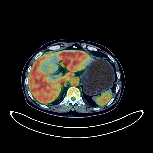
Lung Cancer PET/CT (case 983827-000123 from PETWB-REP)
3 views
9 days ago
Whole-body 18F-FDG PET/CT scan in a patient with Lung Cancer taken from the PETWB-REP dataset. The following English report (translated from original Chinese) is taken verbatim from the public dataset and has not been modified or otherwise checked for accuracy (see the end for citation). Impression a. A large mass in the posterior segment of the left lower lobe, with increased FDG metabolism, suggestive of lung cancer with surrounding inflammation. Multiple lymph node metastases in the left hilum and mediastinum. T7 vertebral metastasis is highly probable. b. Several small chronic inflammatory nodules (solid) in both lungs are highly probable; CT scan for comparison is recommended. A few chronic inflammations and old lesions in both lungs. Some arterial wall calcification. A few ischemic lesions in the deep bilateral brain; age-related brain, MRI is recommended. Small liver cysts. Fatty infiltration of the pancreas. Increased FDG metabolism in some intestinal segments, suggestive of inflammatory or physiological uptake. Degenerative changes in the spine. L4/5 and L5/S1 intervertebral disc bulges. Reactive hyperplasia of bilateral cervical lymph nodes. This case is from PETWB-REP, a curated dataset of whole-body 18F-FDG PET/CT scans and corresponding radiology reports from 490 patients with a broad spectrum of malignancies. The data were retrospectively collected from patients who underwent clinically indicated whole-body 18F-FDG PET/CT scans at the Shanghai Universal Medical Imaging Diagnostic Center between 2021 and 2024. License: Creative Commons Attribution 4.0 International (CC BY 4.0) Citation: Xue, L., Feng, G., Wenbo, Z., Zhang, Y., Li, L., Wang, S., Peng, L., Peng, S., & Gao, X. (2026). PETWB-REP: A Multi-Cancer Whole-Body FDG PET/CT Dataset with Corresponding Radiology Reports [Data set]. Zenodo. https://doi.org/10.5281/zenodo.18670487
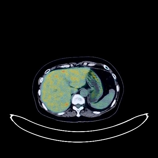
Cervical Cancer PET/CT (case 983827-000005 from PETWB-REP)
2 views
9 days ago
Whole-body 18F-FDG PET/CT scan in a patient with Cervical Cancer taken from the PETWB-REP dataset. The following English report (translated from original Chinese) is taken verbatim from the public dataset and has not been modified or otherwise checked for accuracy (see the end for citation). Impression a. Cervical mass with elevated FDG metabolism, highly suggestive of cervical cancer; please correlate with clinicopathology. b. Multiple uterine fibroids; please correlate with specialist examination. Chronic inflammatory nodules in both lungs; CT follow-up recommended. A few post-inflammatory lesions in both lungs. Calcification of some arterial walls (including coronary arteries). Possible small cyst in the left kidney. Chronic inflammatory changes or physiological uptake in part of the gastric wall and ascending colon; please correlate with endoscopy. Osteoporosis, mild vertebral osteophyte formation, L4/5 and L5/S1 intervertebral disc bulge. No obvious abnormalities seen on cranial scintigraphy. Right upper alveolar alveolitis. Bilateral palatine tonsillitis. This case is from PETWB-REP, a curated dataset of whole-body 18F-FDG PET/CT scans and corresponding radiology reports from 490 patients with a broad spectrum of malignancies. The data were retrospectively collected from patients who underwent clinically indicated whole-body 18F-FDG PET/CT scans at the Shanghai Universal Medical Imaging Diagnostic Center between 2021 and 2024. License: Creative Commons Attribution 4.0 International (CC BY 4.0) Citation: Xue, L., Feng, G., Wenbo, Z., Zhang, Y., Li, L., Wang, S., Peng, L., Peng, S., & Gao, X. (2026). PETWB-REP: A Multi-Cancer Whole-Body FDG PET/CT Dataset with Corresponding Radiology Reports [Data set]. Zenodo. https://doi.org/10.5281/zenodo.18670487
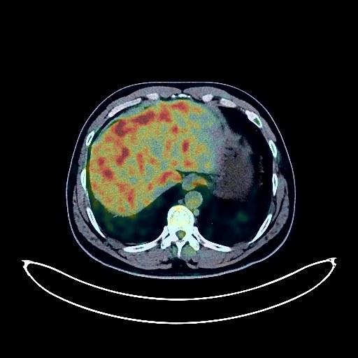
Lung Cancer PET/CT (case 983827-000130 from PETWB-REP)
2 views
9 days ago
Whole-body 18F-FDG PET/CT scan in a patient with Lung Cancer taken from the PETWB-REP dataset. The following English report (translated from original Chinese) is taken verbatim from the public dataset and has not been modified or otherwise checked for accuracy (see the end for citation). Impression a. A mass in the right hilum with increased FDG metabolism, highly suggestive of malignancy, but inflammatory granulomatous lesions need to be ruled out. Please correlate with clinicopathology. b. A solid nodule in the apical segment of the right upper lobe, with normal FDG uptake, suggestive of a chronic inflammatory nodule, metastasis needs to be ruled out. Please monitor with CT. Reactive hyperplasia of mediastinal lymph nodes is highly probable. c. A small chronic inflammatory nodule in the anterior basal segment of the right lower lobe. Emphysema in both upper lobes of the lungs. Right frontal sinusitis, bilateral ethmoid sinusitis, and bilateral maxillary sinusitis. No obvious abnormalities were found on cranial scintigraphy. Continuous increased FDG metabolism in the descending colon and sigmoid colon, highly suggestive of inflammatory uptake or polypoid lesions. Colonoscopy is recommended for clarification. Gallstones. Right kidney stone, small kidney stone in the left kidney. Osteophytes in some vertebrae. This case is from PETWB-REP, a curated dataset of whole-body 18F-FDG PET/CT scans and corresponding radiology reports from 490 patients with a broad spectrum of malignancies. The data were retrospectively collected from patients who underwent clinically indicated whole-body 18F-FDG PET/CT scans at the Shanghai Universal Medical Imaging Diagnostic Center between 2021 and 2024. License: Creative Commons Attribution 4.0 International (CC BY 4.0) Citation: Xue, L., Feng, G., Wenbo, Z., Zhang, Y., Li, L., Wang, S., Peng, L., Peng, S., & Gao, X. (2026). PETWB-REP: A Multi-Cancer Whole-Body FDG PET/CT Dataset with Corresponding Radiology Reports [Data set]. Zenodo. https://doi.org/10.5281/zenodo.18670487
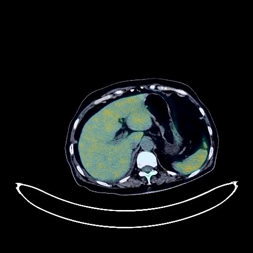
Lung Cancer PET/CT (case 983827-000229 from PETWB-REP)
2 views
9 days ago
Whole-body 18F-FDG PET/CT scan in a patient with Lung Cancer taken from the PETWB-REP dataset. The following English report (translated from original Chinese) is taken verbatim from the public dataset and has not been modified or otherwise checked for accuracy (see the end for citation). Impression a. A soft tissue mass near the hilum of the right upper lobe with increased FDG metabolism, suggestive of malignancy, most likely lung cancer with obstructive changes. Please confirm the diagnosis with pathological examination. Multiple metastatic tumors in the right pleura, right pleural effusion. b. Multiple lymph node metastases in the right hilum and mediastinum. c. Multiple bone metastases throughout the body (see description for details). d. Bilateral adrenal hyperplasia is highly probable; please follow up to rule out metastasis. A few post-inflammatory lesions in both lungs. Partial pulmonary edema in the right lung. Calcification of some arterial walls (including coronary arteries). Calcification in the right lobe of the liver. Small gallstones. Nodular FDG hypermetabolic lesions in the upper abdomen along the course of the small intestine, possibly physiological or inflammatory changes, space-occupying lesion to be ruled out, enhanced CT follow-up recommended. Chronic gastritis changes, gastroscopy follow-up recommended. Spinal degenerative changes. L3/4 intervertebral disc bulge. Bilateral deep cerebral ischemic lesions, mild age-related brain changes, MRI follow-up recommended to rule out micrometastases. Uneven density between the left and right lobes of the thyroid gland; slightly low-density small nodules in the right lobe, FDG metabolism normal, possibly nodular goiter, please follow up with ultrasound. This case is from PETWB-REP, a curated dataset of whole-body 18F-FDG PET/CT scans and corresponding radiology reports from 490 patients with a broad spectrum of malignancies. The data were retrospectively collected from patients who underwent clinically indicated whole-body 18F-FDG PET/CT scans at the Shanghai Universal Medical Imaging Diagnostic Center between 2021 and 2024. License: Creative Commons Attribution 4.0 International (CC BY 4.0) Citation: Xue, L., Feng, G., Wenbo, Z., Zhang, Y., Li, L., Wang, S., Peng, L., Peng, S., & Gao, X. (2026). PETWB-REP: A Multi-Cancer Whole-Body FDG PET/CT Dataset with Corresponding Radiology Reports [Data set]. Zenodo. https://doi.org/10.5281/zenodo.18670487